. 2021 Jul 9;11(1):14181.
doi: 10.1038/s41598-021-93405-3.

A pilot study to assess the healing of meniscal tears in young adult goats

Affiliations

A pilot study to assess the healing of meniscal tears in young adult goats

William Fedje-Johnston et al. Sci Rep. .

Abstract

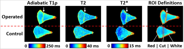
Meniscal tears are a common orthopedic injury, yet their healing is difficult to assess post-operatively. This impedes clinical decisions as the healing status of the meniscus cannot be accurately determined non-invasively. Thus, the objectives of this study were to explore the utility of a goat model and to use quantitative magnetic resonance imaging (MRI) techniques, histology, and biomechanical testing to assess the healing status of surgically induced meniscal tears. Adiabatic T1ρ, T2, and T2* relaxation times were quantified for both operated and control menisci ex vivo. Histology was used to assign healing status, assess compositional elements, and associate healing status with compositional elements. Biomechanical testing determined the failure load of healing lesions. Adiabatic T1ρ, T2, and T2* were able to quantitatively identify different healing states. Histology showed evidence of diminished proteoglycans and increased vascularity in both healed and non-healed menisci with surgically induced tears. Biomechanical results revealed that increased healing (as assessed histologically and on MRI) was associated with greater failure load. Our findings indicate increased healing is associated with greater meniscal strength and decreased signal differences (relative to contralateral controls) on MRI. This indicates that quantitative MRI may be a viable method to assess meniscal tears post-operatively.


Conflict of interest statement

The authors declare no competing interests.

Figures

Figure 1
Figure 1
(A) Excised menisci from the right stifle. Operated meniscus is shown with two arrows pointing to the sutured tears on the cranial aspect (deep/inferior sutures not visible). A singular arrow shows the location of the non-repaired tear on the caudal aspect. This arrow also points in the direction of the main magnetic field (B0) for the MRI experiments. (B) Location of medial meniscal tear incisions shown with dotted lines. Cranial tears were sutured. Caudal tears were left unrepaired. Straight black lines (n = 8) represent approximate MRI imaging planes. Black lines with asterisk represent sectioning locations for histology (n = 4 per goat). Tissue between the straight black lines (3 mm in width circumferentially) was removed for biomechanical testing in the radial direction. The medial meniscus depicted on the left is the contralateral control. (C) Cross section of MRI imaging plane. Meniscal tear shown with dotted line. MRI regions of interest are shown as black (Red ROI), grey (Cut ROI), and white (White ROI).
Figure 2
Figure 2
Quantitative relaxation time maps and region of interest (ROI) definitions for a representative pair of operated and control menisci at the cranial aspect of the menisci and root end of the tear site. The location of the tear is indicated by the arrow. In this example, relaxation times are increased in the vicinity of the tear site, while relaxation times in the white and red zones are similar between the operated and control menisci.
Figure 3
Figure 3
Biomechanical apparatus demonstrating the progression of tissue failure (top). Biomechanical testing of 3 different healing states (bottom): non-healed (blue), healed (red), and control (black).
Figure 4
Figure 4
Comparison of sutured section six weeks post-operative with contralateral control. Serial sections were immunostained with factor VIII-related antigen (top images); the reaction product is brown. Insets demonstrate vessels with endothelial cells positive for factor VIII-related antigen (top left) and area containing no vessels (top right). Complete tissue sections are 1 × objective and insets are 20 × objective. Additional sections were stained with safranin-O (bottom images); red staining indicate the presence of proteoglycans.
Figure 5
Figure 5
Average difference in morphological parameters between induced lesions (sutured/non-sutured) and contralateral control meniscal sections. Vascularity, cell density, proteoglycan content (safranin-O), and type I collagen were binned according to the healing status (non-healed, partially healed, and healed tears). The average difference between non-healed (n = 1), partially healed (n = 4), and healed (n = 3) tears and their respective contralateral control are shown with standard deviation bars. Cell density was the difference in cells per mm2. All other parameters are differences in percent stain/immunostain positivity. Non-healed tear does not have error bars as there was only one tear with this status. Note the dotted black line indicating no difference between induced lesions and their contralateral control.
Figure 6
Figure 6
Relaxation time differences (Δ) between the operated and control menisci. The relaxation time differences were calculated in three ROIs: near the tear site (first column); the white zone side of the tear site (second column); and the red zone side of the tear site (third column). Three relaxation times were evaluated: adiabatic T1ρ (first row); T2 (second row); and T2* (third row). Individual data points are shown as black dots, their mean is plotted as a square, and the error bars show the standard deviation of the mean. Since there was only one non-healed tear, there are no error bars for this point.

References

    1. Fox AJ, Wanivenhaus F, Burge AJ, Warren RF, Rodeo SA. The human meniscus: A review of anatomy, function, injury, and advances in treatment. Clin. Anat. 2015;28:18. doi: 10.1002/ca.22456. - DOI - PubMed
    1. Jorgensen U, Sonne-Holm S, Lauridsen F, Rosenklint A. Long-term follow-up of meniscectomy in athletes. A prospective longitudinal study. J. Bone Joint Surg. B. 1987;69:80–83. doi: 10.1302/0301-620X.69B1.3818740. - DOI - PubMed
    1. Fairbank TJ. Knee joint changes after meniscectomy. J. Bone Joint Surg. 1948;30:664–670. doi: 10.1302/0301-620X.30B4.664. - DOI - PubMed
    1. Mordecai SC, Al-Hadithy N, Ware HE, Gupte CM. Treatment of meniscal tears: An evidence based approach. World J. Orthop. 2014;5:233. doi: 10.5312/wjo.v5.i3.233. - DOI - PMC - PubMed
    1. Nguyen JC, De Smet AA, Graf BK, Rosas HG. MR imaging-based diagnosis and classification of meniscal tears. Radiographics. 2014;34:981. doi: 10.1148/rg.344125202. - DOI - PubMed

Publication types