. 2021 Nov;23(11):2138-2149.
doi: 10.1038/s41436-021-01260-4. Epub 2021 Jul 9.

Biallelic variants in PCDHGC4 cause a novel neurodevelopmental syndrome with progressive microcephaly, seizures, and joint anomalies

Collaborators, Affiliations

Biallelic variants in PCDHGC4 cause a novel neurodevelopmental syndrome with progressive microcephaly, seizures, and joint anomalies

Maria Iqbal et al. Genet Med. 2021 Nov.

Abstract

Purpose: We aimed to define a novel autosomal recessive neurodevelopmental disorder, characterize its clinical features, and identify the underlying genetic cause for this condition.

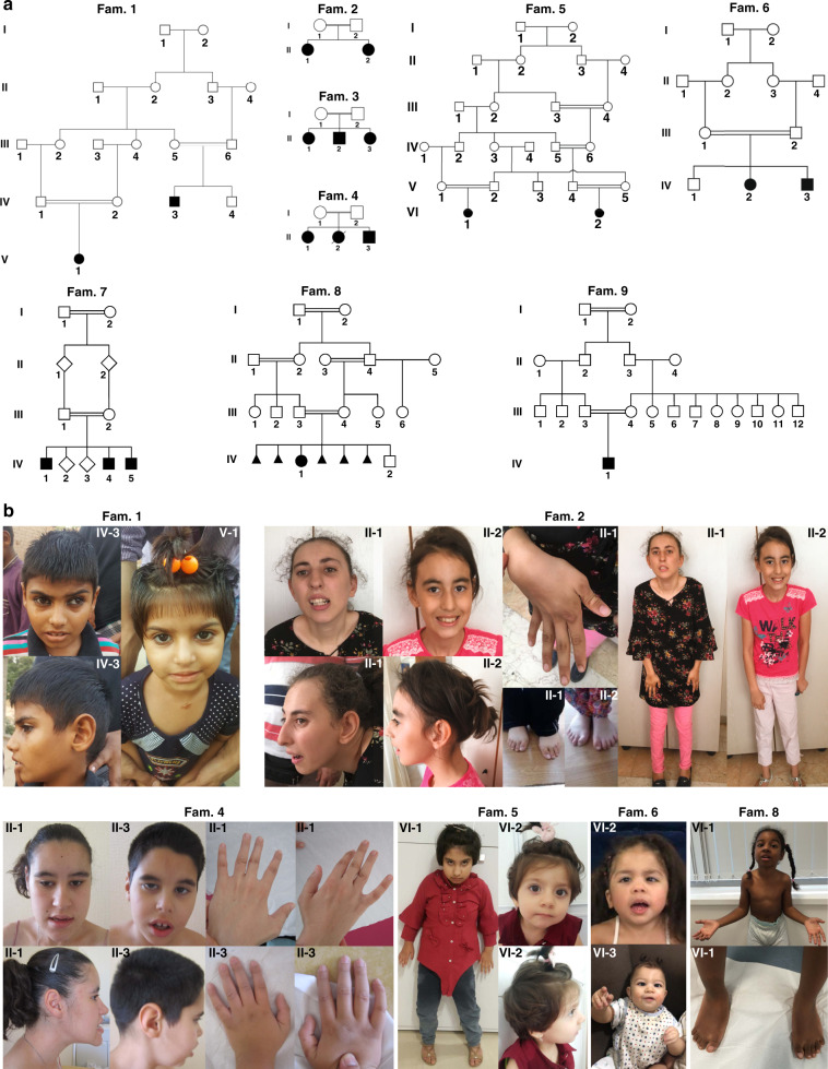
Methods: We performed a detailed clinical characterization of 19 individuals from nine unrelated, consanguineous families with a neurodevelopmental disorder. We used genome/exome sequencing approaches, linkage and cosegregation analyses to identify disease-causing variants, and we performed three-dimensional molecular in silico analysis to predict causality of variants where applicable.

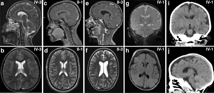
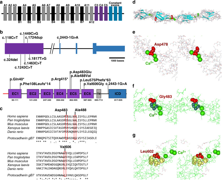
Results: In all affected individuals who presented with a neurodevelopmental syndrome with progressive microcephaly, seizures, and intellectual disability we identified biallelic disease-causing variants in Protocadherin-gamma-C4 (PCDHGC4). Five variants were predicted to induce premature protein truncation leading to a loss of PCDHGC4 function. The three detected missense variants were located in extracellular cadherin (EC) domains EC5 and EC6 of PCDHGC4, and in silico analysis of the affected residues showed that two of these substitutions were predicted to influence the Ca2+-binding affinity, which is essential for multimerization of the protein, whereas the third missense variant directly influenced the cis-dimerization interface of PCDHGC4.

Conclusion: We show that biallelic variants in PCDHGC4 are causing a novel autosomal recessive neurodevelopmental disorder and link PCDHGC4 as a member of the clustered PCDH family to a Mendelian disorder in humans.


Conflict of interest statement

V.K. and C.B. are employees of Centogene GmbH (Rostock, Germany). The other authors declare no competing interests.

Figures

Fig. 1
Fig. 1. Pedigrees and clinical characteristics of individuals harboring biallelic disease-causing variants in PCDHGC4.
(a) Pedigrees of nine unrelated families with disease-causing variants in PCDHGC4. All affected siblings (solid symbols) in each family carry homozygous disease-causing variants in PCDHGC4 while unaffected  parents are heterozygous for identified PCDHGC4 variants (white symbols). (b) Upper panel: facial features of subjects IV-3 and V-1 from family 1 (left), clinical characteristics of subjects II-1 and II-2 from family 2 showing kyphoscoliosis, clinodactyly and hallux valgus (subject II-1), and kyphosis and hypoplasia of the toes (subject II-2). Lower panel (from left to right): facial features and hand anomalies observed in subjects II-1 (22 years) and II-3 (14 years) from family 4, clinical characteristics of subjects VI-1 and VI-2 from family 5, and subjects IV-2 and IV-3 from family 6, and facial features and feet anomalies observed subject VI-1 from family 8.
Fig. 2
Fig. 2. Neuroradiologic features of affected individuals.
Sagittal (a) and axial (b) T2-weighted images of subject IV-3 from family 1 at the age of 10 revealed no structural brain anomalies but showed microcephaly and thin cerebral cortex. (c) Sagittal T1 section after gadolinium injection of subject II-1 (family 4) at 10 years of age and (d) axial T2-weighted images at 16 years revealed no brain-specific abnormality except for a discreet prominent aspect of the lateral ventricles. (e) Sagittal T1 section after gadolinium injection and (f) axial T2-weighted images of subject II-3 (family 4) at 7 years of age revealed a prominent aspect of the lateral ventricles, of the 3rd and, to a milder degree, of the 4th ventricle. (g) Coronal T2-weighted and (h) axial T2-Flair images of subject IV-2 (family 6) at 3 months of age showing normal signal intensity, age-appropriate myelination process and slightly enlarged cerebrospinal fluid (CSF). Coronal (i) and sagittal (j) computed tomography (CT) images of the same subject at the age of 3 years revealing left-sided subcortical hypodensity within left temporal lobe and confirming prominent CSF space.
Fig. 3
Fig. 3. Molecular characterization and in silico analysis of identified disease-causing variants in PCDHGC4.
(a) Schematic representation of the human γ-PCDH cluster. Variable exons of the γ-PCDH A and B subfamilies are shown in gray and black, respectively. Variable exons of the γ-PCDH C subfamily are shown in purple, γ-PCDH constant exons in blue. (b) Schematic representation of the genomic (upper panel) and protein structure (lower panel) of PCDHGC4, and localization of the identified disease-causing variants. Introns are shown by black horizontal line, coding exons by purple and blue bars, noncoding regions of exons by small blue bar (upper panel). Scale bar is referring solely to exons. Protein structure of PCDHGC4 with six extracellular cadherin (EC) repeats (purple), the transmembrane region (gray), and the intracellular domain (ICD, blue). (c) Amino acid sequence alignment of PCDHGC4 across different species including mouse Pcdhgb7 (lower line, all panels) for residues p.Asp483 and p.Ala488 (upper panel) and p.Val606 (lower panel) that are altered in the affected subjects. Protein sequences were prepared from UniProtKB and alignment was performed using Clustal Omega. Position of the altered residues in human are indicated (top numbers). (d) Three-dimensional structure of the EC3 to EC6 domains of Pcdhgb7. Structural information was obtained from the Protein Data Bank (PDB) and is available under the accession number 5v5x. Pcdhgb7 is shown in ribbon representation. β-strands are shown as arrows (blue), a short helical part in red, calcium ions in sphere representation (green), and aspartate at position 478 within the Ca2+-binding DXD motif in space filling representation (red). (e-g) Close up crossed eyes stereo views of p.Asp478 in Pcdhgb7 corresponding to p.Asp483 in PCDHGC4 (e), p.Gly483 (corresponding to p.Ala488 in PCDHGC4) (f), and p.Leu602 (corresponding to p.Val606 in PCDHGC4) (g). Affected amino acid residues are labeled in red, calcium ions are shown in sphere representation (light green), oxygen ligands of the adjacent calcium ion in space filling representation (f, dark green), surrounding hydrophobic residues p.Pro558, p.Tyr604, and p.Val644 of p.Leu602 in space filling representation in yellow (g).

References

    1. Sano K, et al. Protocadherins: a large family of cadherin-related molecules in central nervous system. EMBO J. 1993;12:2249–2256. doi: 10.1002/j.1460-2075.1993.tb05878.x. - DOI - PMC - PubMed
    1. Rubinstein R, et al. Molecular logic of neuronal self-recognition through protocadherin domain interactions. Cell. 2015;163:629–642. doi: 10.1016/j.cell.2015.09.026. - DOI - PMC - PubMed
    1. Hulpiau P, van Roy F. Molecular evolution of the cadherin superfamily. Int J Biochem Cell Biol. 2009;41:349–369. doi: 10.1016/j.biocel.2008.09.027. - DOI - PubMed
    1. Wu Q, Maniatis T. A striking organization of a large family of human neural cadherin-like cell adhesion genes. Cell. 1999;97:779–790. doi: 10.1016/s0092-8674(00)80789-8. - DOI - PubMed
    1. Sugino H, et al. Genomic organization of the family of CNR cadherin genes in mice and humans. Genomics. 2000;63:75–87. doi: 10.1006/geno.1999.6066. - DOI - PubMed

Publication types