Review
. 2022 Jan;20(1):20-34.
doi: 10.1038/s41579-021-00586-9. Epub 2021 Jul 9.

Structure and function of retroviral integrase

Affiliations
Review

Structure and function of retroviral integrase

Goedele N Maertens et al. Nat Rev Microbiol. 2022 Jan.

Abstract

A hallmark of retroviral replication is establishment of the proviral state, wherein a DNA copy of the viral RNA genome is stably incorporated into a host cell chromosome. Integrase is the viral enzyme responsible for the catalytic steps involved in this process, and integrase strand transfer inhibitors are widely used to treat people living with HIV. Over the past decade, a series of X-ray crystallography and cryogenic electron microscopy studies have revealed the structural basis of retroviral DNA integration. A variable number of integrase molecules congregate on viral DNA ends to assemble a conserved intasome core machine that facilitates integration. The structures additionally informed on the modes of integrase inhibitor action and the means by which HIV acquires drug resistance. Recent years have witnessed the development of allosteric integrase inhibitors, a highly promising class of small molecules that antagonize viral morphogenesis. In this Review, we explore recent insights into the organization and mechanism of the retroviral integration machinery and highlight open questions as well as new directions in the field.


Conflict of interest statement

Conflicts of Interest

A.N.E. consults for ViiV Healthcare, Co. The other authors have no conflicts to declare.

Figures

Figure 1.
Figure 1.. Schematic of the retroviral integration process.
The intasome undergoes two enzymatic steps (blue arrows) en route from the initial stable synaptic complex to the post-catalytic strand transfer complex, wherein 3’ vDNA ends are joined to chromosomal DNA. Following intasome disassembly, ligation of the 5’ vDNA ends to the chromosome requires at least three additional enzymatic functions (thick black arrows) provided by host cell factors.
Figure 2.
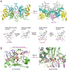
Figure 2.. PFV intasome structures and IN active site mechanics.
(A) Crystal structures of the PFV cleaved synaptic complex (left) and the target capture complex (right),. IN and DNA chains are shown as cartoons and color-coded: target DNA and the scissile vDNA dinucleotides (below) are magenta, the rest of the vDNA is dark grey; IN chains providing the active sites are green and cyan, and the outer IN chains are yellow. Grey spheres are catalytic metal ions. Active sites carboxylates comprising the IN D,D-35-E motif are shown as red sticks. Locations of the individual IN domains (NTD, CCD, and CTD) and the intasome active sites (red arrowheads) are indicated. Bottom panel shows target DNA and/or vDNA within four successive functional states of the intasome as it transitions from the initial stable synaptic complex (left) to the post-catalytic strand transfer complex (right). (B) IN active sites primed for 3’-processing (left) and strand transfer (right). DNA chains and IN active site residues (indicated as D, D, and E) are shown as sticks. DNA chains are coloured dark grey, except for target DNA and scissile vDNA dinucleotide (magenta). Selected water molecules are shown as small red spheres. Direction of the SN2 nucleophilic substitution at each step is indicated with red arrows. Grey and red dashes are hydrogen and metal coordination bonds, respectively.
Figure 3.
Figure 3.. Diversity of retroviral intasome architectures.
Examples of intasome structures from simiispumavirus (PFV, containing a tetramer of IN), δ-retrovirus (STLV, a tetramer), α-retrovirus (ASLV, an octamer), and lentivirus (MVV, a hexadecamer) are shown as cartoons with cylinders representing α-helices,,,. In each case, IN domains contributing to the CIC are shown in colour with the remainder of the structure in grey. Synaptic CTDs in ASLV and MVV intasomes are coloured orange to indicate their origins from flanking IN protomers.
Figure 4.
Figure 4.. Integration into nucleosomal DNA.
Cryo-EM structure of the PFV intasome in a strand transfer complex with a mononucleosome. The structure is shown in two orthogonal orientations. DNA (vDNA and nucleosomal DNA) and protein (IN and histones) are depicted as cartoons or in spacefill, respectively (H2A, orange; H2B, olive; H3 and H4 grey). For clarity, IN chains are hidden in the right panel. Note that the nucleosomal DNA is lifted from the surface of the histone octamer at the site of integration.
Figure 5.
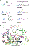
Figure 5.. IN strand transfer inhibitors.
(A) Chemical structures of a lead diketo-acid compound (L-731,988) and of the five INSTIs approved for clinical use. Co-planar oxygen atoms involved in metal chelation are highlighted in red, and halo-benzyl groups in blue. (B) Bictegravir bound to the active site of the red-capped mangabey SIV intasome visualised by cryo-EM. The drug molecule is shown as sticks with carbon atoms in magenta. Note that Asn155 and Gln148 participate in the secondary coordination spheres of the metal ions. In particular, Q148H leads to displacement of a key water molecule (W) bonded to carboxylates of Asp116 and Glu152 (REF.).
Figure 6.
Figure 6.. Allosteric HIV-1 IN inhibitors.
(A) Examples of small molecules of this class. Functional groups comprising the shared ALLINI warhead structure are shown in colour: red, a carboxyl group; green, an aliphatic side chain; and blue, a bulky hydrophobic group. (B) Thin section electron microscopy of stained HIV-1 virions produced under normal conditions (left) or in the presence of an ALLINI (right). Idealised schematics of the respective morphologies are shown on the bottom. (C) Co-crystal structure of HIV-1 IN CCD dimer with GSK1264 (REF.). IN chains are depicted as cartoons, with the amino acid residues lining the ALLINI binding pocket as sticks. Carbon atoms of the inhibitor are shown in magenta. (D) A chain of IN dimers observed in the co-crystal structure of full-length HIV-1 IN with GSK1264 (REF.). Positions of IN CCDs and CTDs are indicated; NTDs were not resolved in this structure. The ALLINI molecule (positions indicated with arrowheads), found at each of the CTD-CCD interfaces, is shown in spacefill, with carbon atoms in magenta.

References

    1. Poiesz BJ et al. Detection and isolation of type C retrovirus particles from fresh and cultured lymphocytes of a patient with cutaneous T-cell lymphoma. Proc Natl Acad Sci U S A 77, 7415–7419, doi:10.1073/pnas.77.12.7415 (1980). - DOI - PMC - PubMed
    1. Coffin JM The discovery of HTLV-1, the first pathogenic human retrovirus. Proc Natl Acad Sci U S A 112, 15525–15529, doi:10.1073/pnas.1521629112 (2015). - DOI - PMC - PubMed
    1. Barre-Sinoussi F et al. Isolation of a T-lymphotropic retrovirus from a patient at risk for acquired immune deficiency syndrome (AIDS). Science 220, 868–871, doi:10.1126/science.6189183 (1983). - DOI - PubMed
    1. Gallo RC et al. Isolation of human T-cell leukemia virus in acquired immune deficiency syndrome (AIDS). Science 220, 865–867, doi:10.1126/science.6601823 (1983). - DOI - PubMed
    1. Baltimore D RNA-dependent DNA polymerase in virions of RNA tumour viruses. Nature 226, 1209–1211, doi:10.1038/2261209a0 (1970). - DOI - PubMed

Publication types