

Editorial
doi: 10.1007/s00424-021-02605-3.
Where vision begins
Affiliations
- PMID: 34245377
- PMCID: PMC8271335
- DOI: 10.1007/s00424-021-02605-3
Item in Clipboard
Editorial
Where vision begins
Pflugers Arch.
2021 Sep.
No abstract available
Figures
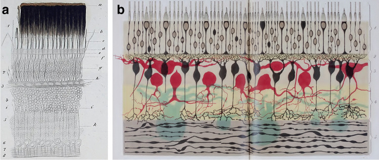
Historical pictures of phototransduction. a Drawing of a vertical section of a fish (perch) retina as published by Heinrich Müller (1856) (original Fig. 1 of Appendix I; book in German) [16]. The sketch shows the principal layers of a vertebrate retina; the pigment epithelium (a, dark brown) covers most of the rod outer segments (1) and parts of the cone outer segments (b). Clearly visible are the outer nuclear layer (2), the outer plexiform layer (3), the inner nuclear layer (4), a further “granular layer,” probably the inner plexiform layer (5), the ganglion cell layer (6, “nerve cell layer”), and the optic nerve fiber layer (7) (b). The first diagram of the retina published by Ferruccio Tartuferi in 1887 (Table XIX; paper in Italian) [20]. The numbers identify correctly the organization of the retinal layers and cells: 1, rod and cone photoreceptors, joined to the outer nuclear layer (“neuroepithelial layer” in the original figure legend); 2, outer plexiform layer (“subepithelial layer”); 3, inner nuclear layer, with horizontal and bipolar cells (“cellular portion of the first cerebral layer” endowed with “plumed cells,” in black, and with “large superficial cells,” in red); 4, inner plexiform layer (“inner reticular layer”); 5, ganglion cell layer, with ganglion cell axons (“layer of the nervous cells,” in light blue, “and of the nerve fibers”)

The membrane ion transport and the phototransduction cascade shared by retinal rods and cones across species. Besides the ubiquitous sustained outward K+ current, many other conductances that have been found in rod and cones of different species are omitted. Other regulatory proteins and pathways, as RD3, the Ca2+-regulation of other enzymes besides GCAP and recoverin (REC), and the pigment epithelium processes that resynthesize Rh, are omitted as well for simplicity and/or because are too speculative yet
Similar articles
-
Vision: from photon to perception.Proc Natl Acad Sci U S A. 1996 Jan 23;93(2):557-9. doi: 10.1073/pnas.93.2.557. Proc Natl Acad Sci U S A. 1996. PMID: 9254392 Free PMC article. No abstract available.
-
[Modulation of visual information in the retina].Sheng Li Ke Xue Jin Zhan. 1991 Jul;22(3):207-11. Sheng Li Ke Xue Jin Zhan. 1991. PMID: 1947980 Review. Chinese. No abstract available.
-
The psychological septum. An investigation into its function.Am J Optom Physiol Opt. 1982 Aug;59(8):639-41. doi: 10.1097/00006324-198208000-00004. Am J Optom Physiol Opt. 1982. PMID: 7137303
-
The movies in our eyes.Sci Am. 2007 Apr;296(4):72-9. doi: 10.1038/scientificamerican0407-72. Sci Am. 2007. PMID: 17479633 No abstract available.
-
What can mice tell us about how vision works?Trends Neurosci. 2011 Sep;34(9):464-73. doi: 10.1016/j.tins.2011.07.002. Epub 2011 Aug 15. Trends Neurosci. 2011. PMID: 21840069 Free PMC article. Review.
Cited by
-
A cytoplasmic protein kinase couples engagement of Chlamydomonas ciliary receptors to cAMP-dependent cellular responses.J Cell Sci. 2022 May 15;135(10):jcs259814. doi: 10.1242/jcs.259814. Epub 2022 May 23. J Cell Sci. 2022. PMID: 35502650 Free PMC article.
-
New tricks and emerging applications from contemporary azobenzene research.Photochem Photobiol Sci. 2022 Oct;21(10):1719-1734. doi: 10.1007/s43630-022-00262-8. Epub 2022 Jul 27. Photochem Photobiol Sci. 2022. PMID: 35896915
-
Light induces a rapid increase in cAMP and activates PKA in rod outer segments of the frog retina.J Gen Physiol. 2024 Nov 4;156(11):e202313530. doi: 10.1085/jgp.202313530. Epub 2024 Oct 22. J Gen Physiol. 2024. PMID: 39436404 Free PMC article.
-
Inherited Retinal Degeneration: Towards the Development of a Combination Therapy Targeting Histone Deacetylase, Poly (ADP-Ribose) Polymerase, and Calpain.Biomolecules. 2023 Mar 23;13(4):581. doi: 10.3390/biom13040581. Biomolecules. 2023. PMID: 37189329 Free PMC article.
References
Publication types
MeSH terms
LinkOut - more resources
Full Text Sources
