. 2021 Aug;35(8):e21775.
doi: 10.1096/fj.202100686R.

Elucidating the structure and functions of Resolvin D6 isomers on nerve regeneration with a distinctive trigeminal transcriptome

Affiliations

Elucidating the structure and functions of Resolvin D6 isomers on nerve regeneration with a distinctive trigeminal transcriptome

Thang L Pham et al. FASEB J. 2021 Aug.

Abstract

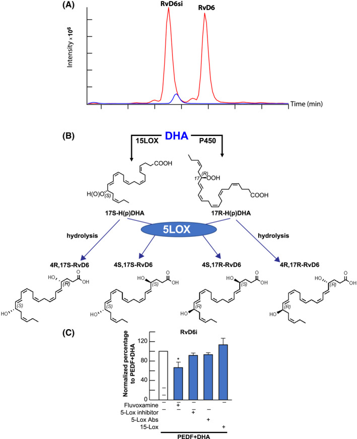
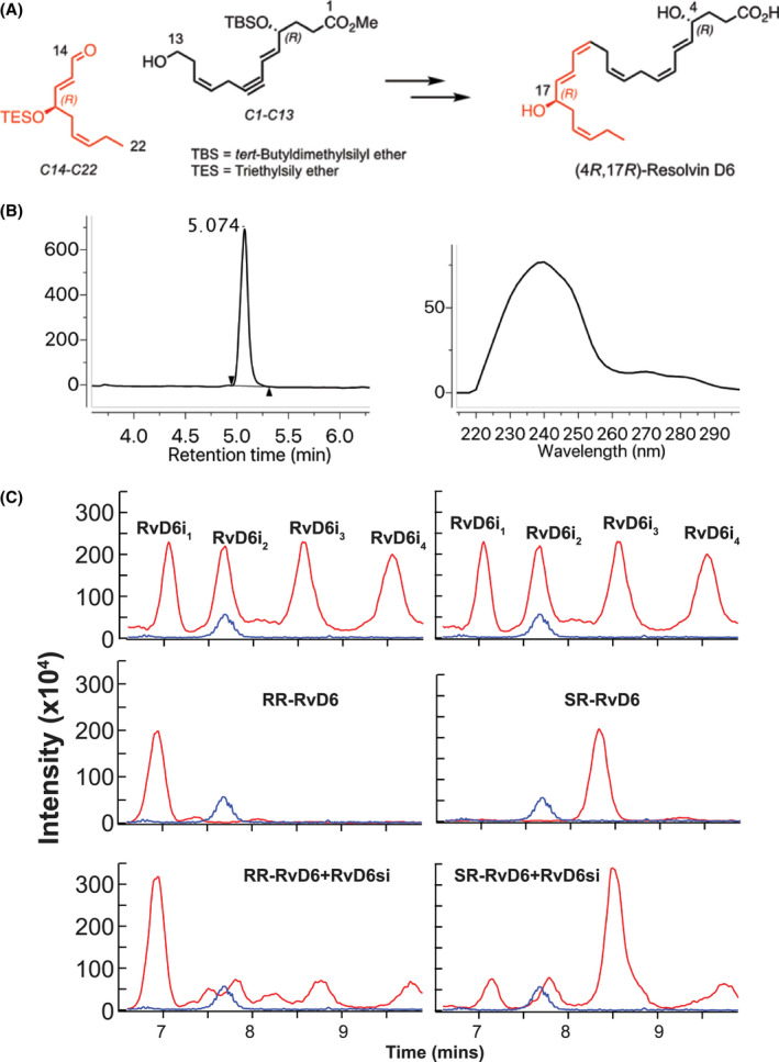
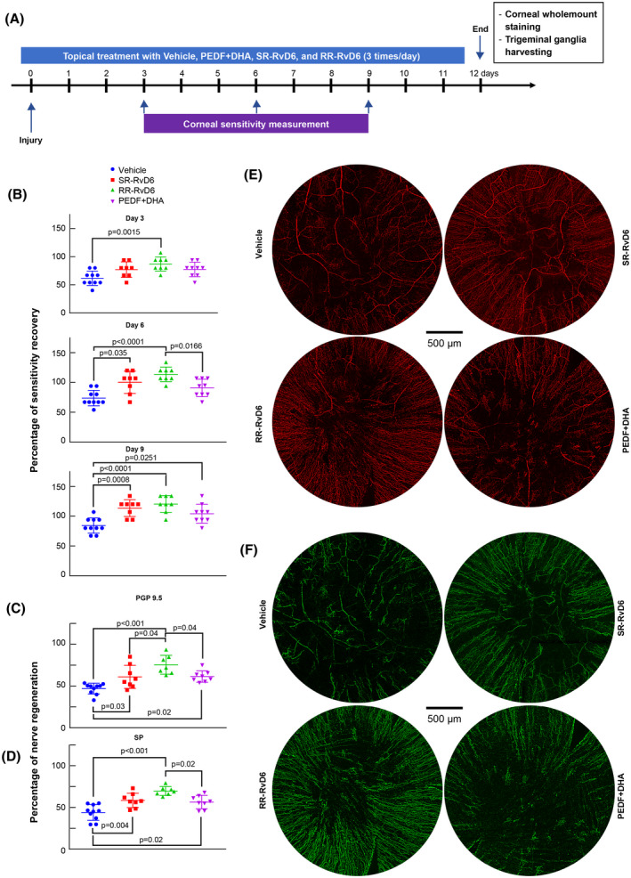
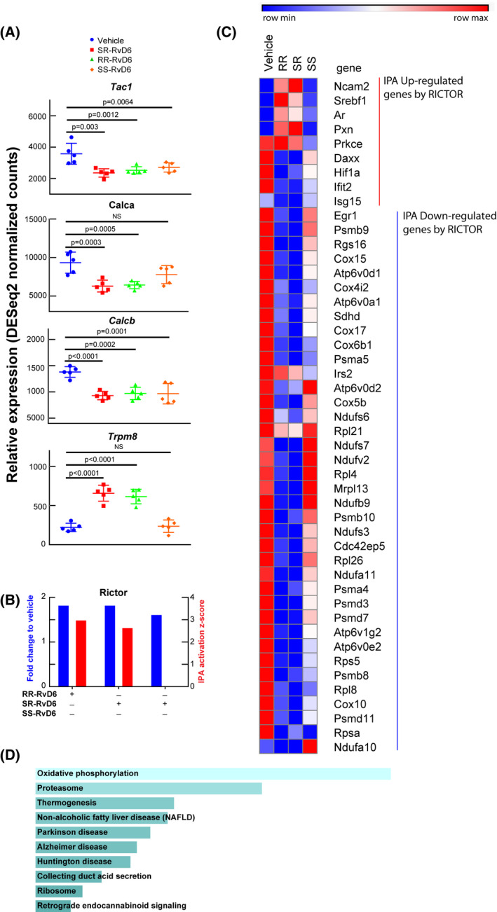
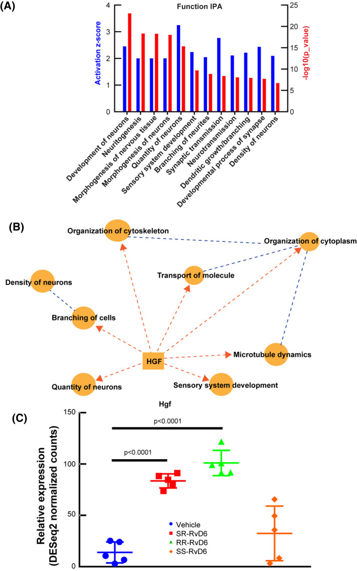
Innervation sustains cornea integrity. Pigment epithelium-derived factor (PEDF) plus docosahexaenoic acid (DHA) regenerated damaged nerves by stimulating the synthesis of a new stereoisomer of Resolvin D6 (RvD6si). Here, we resolved the structure of this lipid isolated from mouse tears after injured corneas were treated with PEDF + DHA. RvD6si synthesis was inhibited by fluvoxamine, a cytochrome P450 inhibitor, but not by 15- or 5-LOX inhibitors, suggesting that the 4- and 17-hydroxy of DHA have an RR- or SR-configuration. The two compounds were chemically synthesized. Using chiral phase HPLC, four peaks of RvD6si1-4 from tears were resolved. The RR-RvD6 standard eluted as a single peak with RvD61 while pure SR-RvD6 eluted with RvD63 . The addition of these pure mediators prompted a trigeminal ganglion transcriptome response in injured corneas and showed that RR-RvD6 was the more potent, increasing cornea sensitivity and nerve regeneration. RR-RvD6 stimulates Rictor and hepatocyte growth factor (hgf) genes specifically as upstream regulators and a gene network involved in axon growth and suppression of neuropathic pain, indicating a novel function of this lipid mediator to maintain cornea integrity and homeostasis after injury.

Keywords: Rictor; RvD6 stereoisomers; corneal nerves; hgf; pain; trigeminal ganglia transcriptome.


Conflict of interest statement

The subject matter of this manuscript is protected by a US provisional patent application assigned to the Board of Supervisors of Louisiana State University and Agricultural and Mechanical College (LSU) and licensed to a company, CurVirBiotech, founded by co‐author Nicolas G. Bazan.

Figures

FIGURE 1
FIGURE 1
Effect of inhibitors on RvD6si synthesis. A, Different retention time of standard RvD6 (SS‐RvD6) and the RvD6si isomer isolated from tears of PEDF + DHA‐treated mice. The blue peak represents the internal standard LTB4‐d4. The RvD6si eluted earlier than LTB4‐d4 and RvD6 standard. B, Possible stereoisomers of RvD6 made from DHA. The 15‐LOX derivatives of RvD6 (left panel) contain the 17S‐OH, while the P450 derivatives (right panel) contain the 17R‐OH on the DHA backbone. 5‐LOX catalyzes the formation of 4S‐OH, while the 4R‐OH might be formed by hydrolysis. C, Incubating corneas with inhibitors/antibodies in the presence of PEDF + DHA shows that only the cytochrome P450 inhibitor Fluvoxamine, significantly reduced the synthesis of RvD6si while 15‐ and 5‐LOX inhibitors/antibodies had no effect. Details about inhibitors and antibodies used are described in Table 1. Values are mean ± SD * P < .05 using Student's t test with PEDF + DHA group as reference. There were 10 corneas/sample per experiment (total 50 corneas for 5 groups of treatment). An average of three independent experiments was calculated
FIGURE 2
FIGURE 2
The structure of RvD6si was confirmed by total synthesis. A, The synthetic scheme of RR‐RvD6 involved key synthetic intermediates C1‐C13 (black) and C14‐C22 (red) were constructed from chirally pure building blocks that were synthesized in conjunction with stereospecific transformations to furnish the target with absolute stereo‐ and regiochemical control. B. The purity of RR‐RvD6 was confirmed by LC‐MS chromatogram (left) while its characteristic conjugated 1,3‐butadiene moieties are shown in the molecule at carbon positions C5‐C7 and C13‐C15, confirmed by UV spectrum (right). B, LC‐MS/MS analysis of mouse tears‐derived RvD6si using a chiral column shows the separation of four peaks. Chemical synthetized RR‐RvD6 had the same retention time as the first peak of RvD6si, while the SR‐RvD6 partially matched the third peak of RvD6 isomer. Only RR‐RvD6 has an earlier retention time than the internal standard LTB4‐d4 (blue), as shown in Figure 1A
FIGURE 3
FIGURE 3
Sensitivity and corneal nerve regeneration by the two RvD6 isomers. A, Experimental design to determine the biological activities of pure RR‐RvD6 and SR‐RvD6. The vehicle was used as negative control while the PEDF + DHA was used as positive reference. B, Corneal sensitivity was recorded with a non‐contact corneal esthesiometer every 3 days after injury. RR‐RvD6‐treated mice show faster sensation recovery at day 3, followed by SR‐RvD6 at day 6, and PEDF + DHA at day 9 after injury and treatment. The vehicle is used as comparative reference. Nerve density and representative whole‐mount images of PGP 9.5 (C, E) and SP (D, F) positive axons at 12 days after injury and treatment. All data were normalized to the baseline (uninjured corneas) as 100%. The p values were calculated by one‐way ANOVA, followed by Tukey's honest significant difference (HSD) multiple pairwise comparisons
FIGURE 4
FIGURE 4
Transcriptome signature in the trigeminal ganglion unravels the role of RvD6 isomers. A, RNA‐seq normalized counts of genes involved in inflammation and pain, including Tac1, Calca, Calcb, and Trpm8. The p values are derived from ANOVA post hoc Dunnett's multiple comparisons test with vehicle as a reference. Mean and SD are depicted as the lines. Each data point represents an ipsilateral TG of a mouse. B, The Rictor signaling activated by RvD6 isomers. The blue columns represent the relative normalized fold changes of Rictor gene analyzed by RNA‐seq, while the red columns represent the activation z‐score of Rictor signaling predicted by IPA using RNA‐seq data as input. The SS‐RvD6 z‐score was insufficient to show the activation of RICTOR pathway in the IPA. C, The RNA‐seq normalized counts heatmap of RICTOR regulated genes. The genes activated (red) and inhibited (blue) are grouped and clustered across all treatment groups. There are about 5 upregulated genes and 36 downregulated genes by SR‐and RR‐RvD6. D, The KEGG‐pathway prediction of RICTOR downregulated genes from C. Bars were sorted by p value. The length of the bar represents the significance of the pathway, while the lighter the color, the higher the significance
FIGURE 5
FIGURE 5
Predicted action of RR‐RvD6 on nerve growth in the trigeminal ganglia. A, The IPA of nervous system development and functions in the TG after RR‐RvD6‐treated corneas in mice. The bar chart shows the activation z‐score (blue) and p value (red) of 12 significant groups of biological functions. B, The graphical summary of RR‐RvD6 induced axon regeneration with hgf expression as a central upstream modulator. Dash lines represent that Hgf requires several intermediate signaling pathways to reach the biological function. C, Hgf was found significantly elevated by S R and RR‐RvD6 treatment but not by SS‐RvD6

References

    1. Müller LJ, Marfurt CF, Kruse F, Tervo TMT. Corneal nerves: structure, contents and function. Exp Eye Res. 2003;76(5):521‐542. - PubMed
    1. He J, Bazan HEP. Mapping the nerve architecture of diabetic human corneas. Ophthalmology. 2012;119(5):956‐964. 10.1016/j.ophtha.2011.10.036 - DOI - PMC - PubMed
    1. Hamrah P, Cruzat A, Dastjerdi MH, et al. Corneal sensation and subbasal nerve alterations in patients with herpes simplex keratitis: an in vivo confocal microscopy study. Ophthalmology. 2010;117(10):1930‐1936. 10.1016/j.ophtha.2010.07.010 - DOI - PMC - PubMed
    1. Cruzat A, Witkin D, Baniasadi N, et al. Inflammation and the nervous system: the connection in the cornea in patients with infectious keratitis. Invest Ophthalmol Vis Sci. 2011;52(8):5136‐5143. 10.1167/iovs.10-7048 - DOI - PMC - PubMed
    1. He J, Bazan HEP. Corneal nerve architecture in a donor with unilateral epithelial basement membrane dystrophy. Ophthalmic Res. 2013;49(4):185‐191. 10.1159/000345766 - DOI - PMC - PubMed

Publication types