. 2021 Sep 15:48:128252.
doi: 10.1016/j.bmcl.2021.128252. Epub 2021 Jul 7.

A microtubule-localizing activity-based sensing fluorescent probe for imaging hydrogen peroxide in living cells

Affiliations

A microtubule-localizing activity-based sensing fluorescent probe for imaging hydrogen peroxide in living cells

Shang Jia et al. Bioorg Med Chem Lett. .

Abstract

Hydrogen peroxide (H2O2) is a major reactive oxygen species (ROS) in living systems with broad roles spanning both oxidative stress and redox signaling. Indeed, owing to its potent redox activity, regulating local sites of H2O2 generation and trafficking is critical to determining downstream physiological and/or pathological consequences. We now report the design, synthesis, and biological evaluation of Microtubule Peroxy Yellow 1 (MT-PY1), an activity-based sensing fluorescent probe bearing a microtubule-targeting moiety for detection of H2O2 in living cells. MT-PY1 utilizes a boronate trigger to show a selective and robust turn-on response to H2O2 in aqueous solution and in living cells. Live-cell microscopy experiments establish that the probe co-localizes with microtubules and retains its localization after responding to changes in levels of H2O2, including detection of endogenous H2O2 fluxes produced upon growth factor stimulation. This work adds to the arsenal of activity-based sensing probes for biological analytes that enable selective molecular imaging with subcellular resolution.

Keywords: Activity-based sensing; Fluorescent probe; Hydrogen peroxide; Molecular imaging; Reactive oxygen species.


Conflict of interest statement

Declaration of Competing Interest

The authors declare that they have no known competing financial interests or personal relationships that could have appeared to influence the work reported in this paper.

Figures

Figure 1.
Figure 1.
Structures of (a) microtubule-targeting molecule docetaxel; (b) mitochondria-localizing H2O2 probe Mito-PY1, and (c) microtubule-localizing H2O2 probe MT-PY1.
Figure 2.
Figure 2.
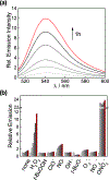
In vitro characterization of MT-PY1. (a) Fluorescence emission spectrum of 5 μM MT-PY1 in 25 mM HEPES buffer pH 7.4 (bottom), and its turn-on response after treatment with 100 μM H2O2 at 37 °C for 5, 15, 30, 45 and 60 min. (b) Fluorescence responses of 5 μM MT-PY1 to 100 μM of various reactive oxygen and nitrogen species at 37 °C after 5, 15, 30, 45 and 60 min of incubation.
Figure 3.
Figure 3.
Confocal microscopy images of MT-PY1 localization in HeLa cells. Cells were incubated with 1 μM MT-PY1 for 15 min at 37 °C prior to imaging. (a) A HeLa cell in metaphase. (b) A HeLa cell in interphase. Cell nucleus is stained by Hoechst 33342 in (a) and (b). (c) HeLa cells expressing mCherry-tubulin stained with MT-PY1. Enlarged image is shown in Supplementary Figure S2. Scale-bar: 10 μm.
Figure 4.
Figure 4.
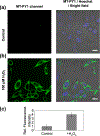
Fluorescence responses of MT-PY1 to exogenous addition of H2O2 in living cells. HeLa cells were incubated with 1 μM MT-PY1 for 0.5 h at 37 °C, followed by treatment with (a) vehicle control or (b) 100 μM H2O2 at 37 °C for 0.5 h; quantification is shown in (c). Scale-bar: 20 μm.
Figure 5.
Figure 5.
Fluorescence imaging of endogenous H2O2 generated by the EGF signaling pathway in live A431 cells. A431 cells incubated with 1 μM MT-PY1 for 0.5 h at 37 °C were treated with (a) vehicle control, (b) 100 ng/mL EGF, (c) 100 ng/mL EGF and 500 μM L-NAME or (d) 100 ng/mL EGF and 50 μM PD15305 for 30 min at 37 °C and imaged. (e) Zoomed-in images of A431 stained with MT-PY1 showing the high spatial resolution of this fluorescence probe. (f) Quantification of fluorescence intensity of a-d shown as mean ± s.d. Scale-bar: 50 μm.
Scheme 1.
Scheme 1.
Synthesis of MT-PY1 and its fluorescence turn-on reaction upon activity-based sensing of H2O2.

References

    1. Snezhkina AV, Kudryavtseva AV, Kardymon OL, et al. ROS generation and antioxidant defense systems in normal and malignant cells. Oxid Med Cell Longev. 2019;2019:1–17. 10.1155/2019/6175804. - DOI - PMC - PubMed
    1. Schieber M, Chandel NS. ROS function in redox signaling and oxidative stress. Curr Biol. 2014;24(10):R453–R462. 10.1016/j.cub.2014.03.034. - DOI - PMC - PubMed
    1. Lisanti MP, Martinez-Outschoorn UE, Lin Z, et al. Hydrogen peroxide fuels aging, inflammation, cancer metabolism and metastasis. Cell Cycle. 2011;10(15): 2440–2449. 10.4161/cc.10.15.16870. - DOI - PMC - PubMed
    1. Ishikawa K, Takenaga K, Akimoto M, et al. ROS-generating mitochondrial DNA mutations can regulate tumor cell metastasis. Science. 2008;320(5876):661–664. 10.1126/science:1156906. - DOI - PubMed
    1. Chandel NS, Vander Heiden MG, Thompson CB, Schumacker PT. Redox regulation of P53 during hypoxia. Oncogene. 2000;19(34):3840–3848. 10.1038/sj.onc.1203727. - DOI - PubMed

Publication types