Review
. 2021 Sep:167:203716.
doi: 10.1016/j.cdev.2021.203716. Epub 2021 Jul 7.

Conditional specification of endomesoderm

Affiliations
Review

Conditional specification of endomesoderm

David R McClay et al. Cells Dev. 2021 Sep.

Abstract

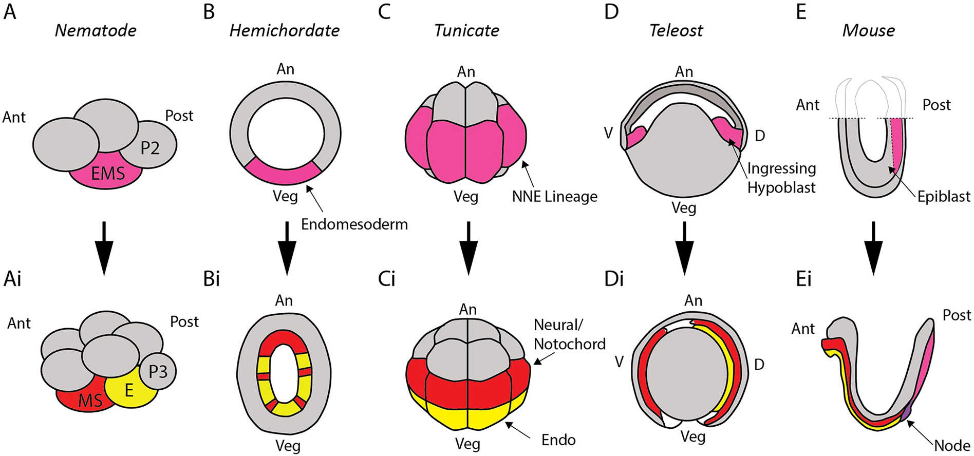
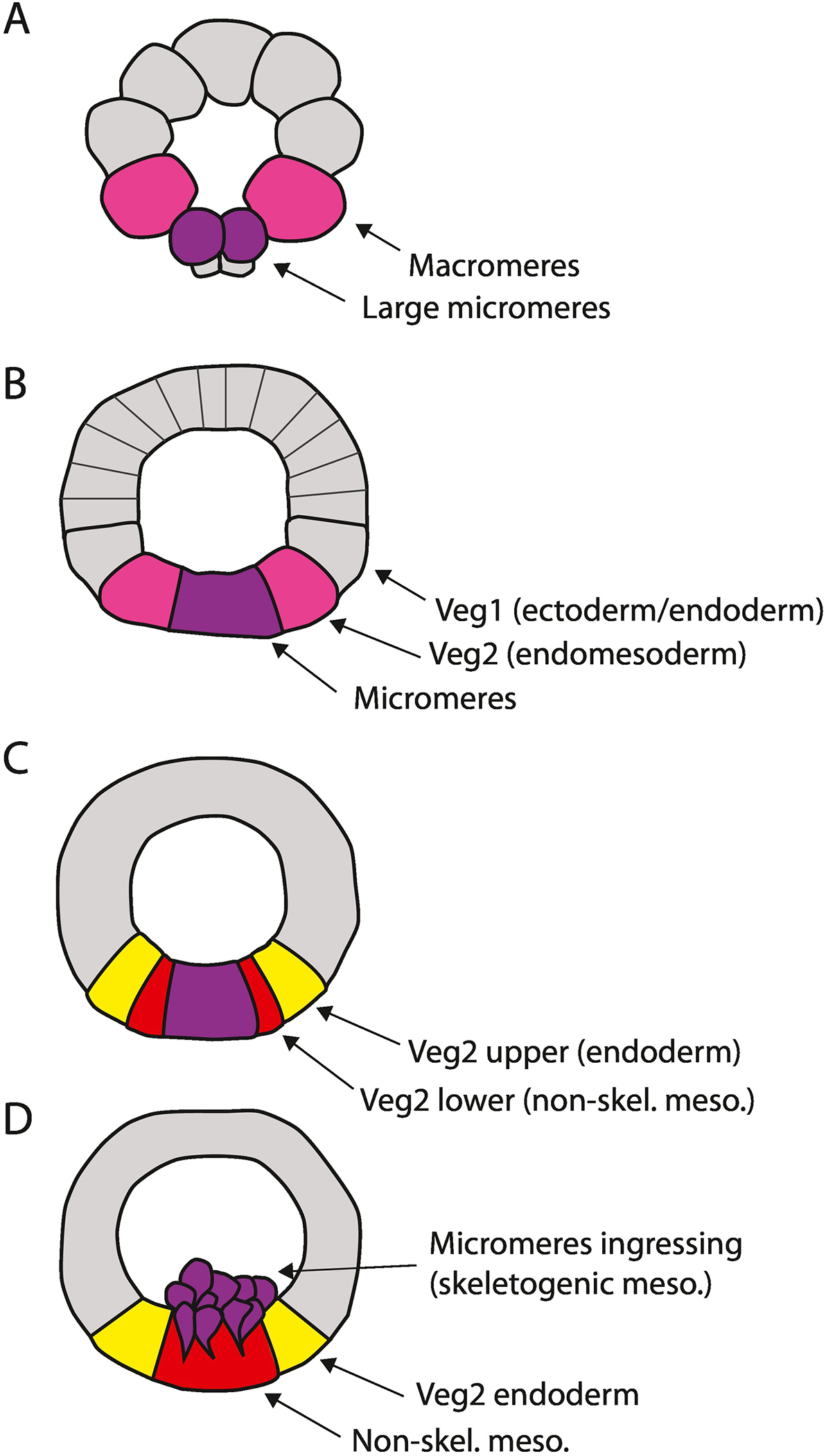
Early in animal development many cells are conditionally specified based on observations that those cells can be directed toward alternate fates. The endomesoderm is so named because early specification produces cells that often have been observed to simultaneously express both early endoderm and mesoderm transcription factors. Experiments with these cells demonstrate that their progeny can directed entirely toward endoderm or mesoderm, whereas normally they establish both germ layers. This review examines the mechanisms that initiate the conditional endomesoderm state, its metastability, and the mechanisms that resolve that state into definitive endoderm and mesoderm.

Keywords: Conditional specification; Endoderm; Frog; Hemichordate; Mesoderm; Nematode; Regulative development; Sea urchin; Tunicate; Zebrafish.


Figures

Figure 1:
Figure 1:
Conditionally specified endomesoderm (or mesendoderm) shown in magenta across a selection of metazoan embryos as it resolves into endodermal lineages (yellow) and mesodermal lineages (red). In nematode embryos (A), the EMS cell gives rise to the MS and E cells (Ai), which specify mesoderm and endoderm respectively. In developing hemichordates (B), a conditionally specified endomesoderm later resolves into endoderm and mesoderm after gastrulation (Bi). Tunicate embryos at the 16-cell stage (C) exhibit high levels of nuclear β-catenin in the NNE lineage before it resolves into endoderm and neural-notochord (i.e. ectoderm-mesoderm) lineages (Ci). In fish embryos (D), during epiboly, ingressing hypoblast cells give rise to either endoderm or mesoderm (Di), suggesting a conditionally specified fate earlier. Mouse embryo epiblast (E) also exhibits cells with a potential to form endoderm or mesoderm (Ei), fates that become committed as cells pass through the primitive streak.
Figure 2:
Figure 2:
Conditional specification of endomesoderm in the sea urchin. At the 32-cell stage (A) the large micromeres (purple) first activate delta expression. The neighboring macromeres (magenta) display a high level of nuclear β-catenin that, along with the Delta input, initiates a conditionally specified endomesoderm. These signals persist through at least the 8th cleavage. (C) Eighth cleavage divides Veg2 cells into two tiers. The anterior progeny (the Veg2 upper tier) lose contact with the micromeres and therefore with the Delta signal thereby losing their mesodermal potential, while the Veg2 lower cells remain in contact with the Delta producing micromeres and adopt a mesodermal fate. (D) At the beginning of gastrulation, the micromeres undergo an epithelial-mesenchymal transition to become the PMCs, and the non-skeletal mesoderm will soon initiate invagination of the archenteron, followed by invagination of the Veg2 endoderm and finally the Veg1 endoderm.
Figure 3:
Figure 3:
Delta-Notch signaling from the micromeres establishes mesoderm. Micromeres first appear at the 16-cell stage (A), activate delta expression at the 32-cell stage and begin expressing Delta at the 60-cell stage (Croce and McClay, 2010). (Ai) This signaling induces mesoderm gene expression in the adjacent endomesoderm (Veg2) cell layer (Ransick et al., 2002; Croce and McClay, 2010; Peter and Davidson, 2010). (Aii) Gcm, a mesoderm marker in the endomesoderm, is expressed at hatched blastula (HB) (reproduced from Croce and McClay, 2010). (B) If the micromeres are surgically removed at the 16-cell stage all endomesoderm is specified as endoderm only (Bi) because Delta is absent. (Bii) Consequently gcm is not expressed. (C) A single labeled micromere reintroduced at 16-cell stage in an otherwise micromere deficient embryo is sufficient to rescue mesoderm development and gcm expression (Ci, Cii). (D) If a micromere from a 16-cell stage embryo is transplanted to an ectopic location of a 60-cell stage wild-type embryo between the Veg1 and Veg2 layers that ectopic micromere induces ectopic mesoderm in the adjacent Veg2 lower and Veg2 upper tissues (Di, Dii). Embryos Aii, Bii, Cii and Dii are at the HB stage (8 to 9 hours post-fertilization). They are all in lateral view with the animal pole to the top, except the embryo in Dii that is rotated to view the embryo from the vegetal pole to show the ring of mesoderm expressing gcm surrounding the endogenous and ectopic micromeres, which otherwise express T-brain, a micromere-descendant marker.

References

    1. Acloque H, Ocana OH, Matheu A, Rizzoti K, Wise C, Lovell-Badge R, Nieto MA, 2011. Reciprocal repression between Sox3 and snail transcription factors defines embryonic territories at gastrulation. Dev Cell 21, 546–558. - PMC - PubMed
    1. Cano A, Perez-Moreno MA, Rodrigo I, Locascio A, Blanco MJ, del Barrio MG, Portillo F, Nieto MA, 2000. The transcription factor snail controls epithelial-mesenchymal transitions by repressing E-cadherin expression. Nat Cell Biol 2, 76–83. - PubMed
    1. Charney RM, Paraiso KD, Blitz IL, Cho KWY, 2017. A gene regulatory program controlling early Xenopus mesendoderm formation: Network conservation and motifs. Semin Cell Dev Biol 66, 12–24. - PMC - PubMed
    1. Cheng X, Lyons DC, Socolar JE, McClay DR, 2014. Delayed transition to new cell fates during cellular reprogramming. Dev Biol 391, 147–157. - PMC - PubMed
    1. Chiu WT, Charney Le R, Blitz IL, Fish MB, Li Y, Biesinger J, Xie X, Cho KW, 2014. Genome-wide view of TGFbeta/Foxh1 regulation of the early mesendoderm program. Development 141, 4537–4547. - PMC - PubMed

Publication types

LinkOut - more resources