. 2021 Nov;100(5):1037-1053.
doi: 10.1016/j.kint.2021.06.031. Epub 2021 Jul 8.

The ketone body β-hydroxybutyrate mitigates the senescence response of glomerular podocytes to diabetic insults

Affiliations

The ketone body β-hydroxybutyrate mitigates the senescence response of glomerular podocytes to diabetic insults

Yudong Fang et al. Kidney Int. 2021 Nov.

Erratum in

Abstract

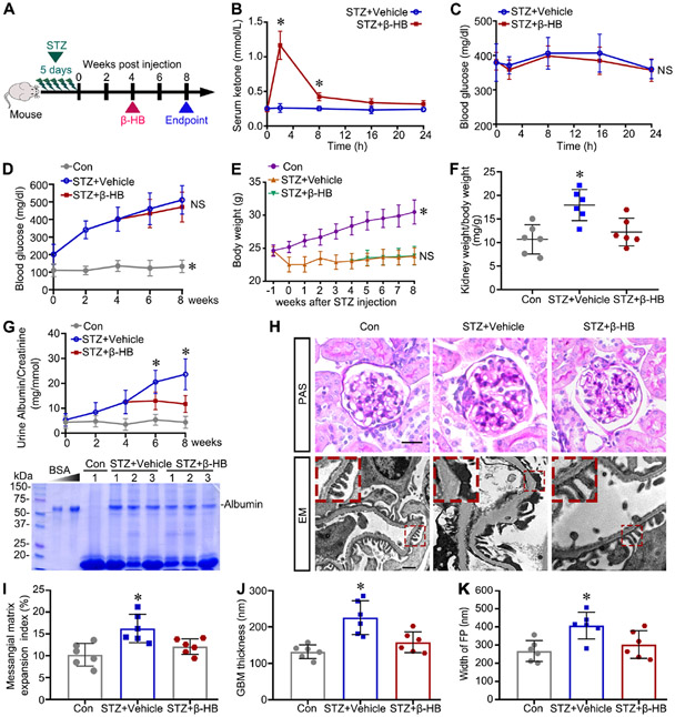
Diabetic kidney disease (DKD) is one of the most common complications of diabetes and is clinically featured by progressive albuminuria, consequent to glomerular destruction that involves podocyte senescence. Burgeoning evidence suggests that ketosis, in particular β-hydroxybutyrate, exerts a beneficial effect on aging and on myriad metabolic or chronic diseases, including obesity, diabetes and chronic kidney diseases. Its effect on DKD is largely unknown. In vitro in podocytes exposed to a diabetic milieu, β-hydroxybutyrate treatment substantially mitigated cellular senescence and injury, as evidenced by reduced formation of γH2AX foci, reduced staining for senescence-associated-β-galactosidase activity, diminished expression of key mediators of senescence signaling like p16INK4A and p21, and preserved expression of synaptopodin. This beneficial action of β-hydroxybutyrate coincided with a reinforced transcription factor Nrf2 antioxidant response. Mechanistically, β-hydroxybutyrate inhibition of glycogen synthase kinase 3β (GSK3β), a convergent point for myriad signaling pathways regulating Nrf2 activity, seems to contribute. Indeed, trigonelline, a selective inhibitor of Nrf2, or ectopic expression of constitutively active mutant GSK3β abolished, whereas selective activation of Nrf2 was sufficient for the anti-senescent and podocyte protective effects of β-hydroxybutyrate. Moreover, molecular modeling and docking analysis revealed that β-hydroxybutyrate is able to directly target the ATP-binding pocket of GSK3β and thereby block its kinase activity. In murine models of streptozotocin-elicited DKD, β-hydroxybutyrate therapy inhibited GSK3β and reinforced Nrf2 activation in glomerular podocytes, resulting in lessened podocyte senescence and injury and improved diabetic glomerulopathy and albuminuria. Thus, our findings may pave the way for developing a β-hydroxybutyrate-based novel approach of therapeutic ketosis for treating DKD.

Keywords: GSK3β; Nrf2 antioxidant response; aging; diabetic nephropathy; intermittent fasting; ketosis; time-restricted feeding.


Conflict of interest statement

DISCLOSURE

All the authors declared no competing interests.

Figures

Figure 1.
Figure 1.. β-Hydroxybutyrate prevents the diabetic milieu-induced senescence and injury in cultured podocytes.
Differentiated immortalized murine podocytes were exposed to control medium that contained normal podocyte culture medium consisting of 5mM glucose and 20 mM mannitol as osmotic control (Con), or a diabetic milieu consisting of high glucose (25 mM; HG) and TGFβ1 (2 ng/ml) with or without β-Hydroxybutyrate (β-HB, 4 mM) for 48 hours. (A) Representative light micrographs of podocyte morphology at 48-hour and SA-β-gal staining are shown with typical positive staining being indicated by arrows. Scale bar, 50 μm. (B) Absolute counting of the SA-β-gal-positive cells as percentage of the total number of podocytes per high-power field (n = 3). *P < 0.05 versus other groups. (C) Cells were fixed and subjected to immunofluorescence staining for γH2AX (green) followed by counterstaining with 4′,6-diamidino-2-phenylindole (DAPI) for nuclei or with rhodamine phalloidin for F-actin (red). Representative images of laser scanning confocal fluorescence microscopy are shown. Scale bar, 20 μm. (D) Cell lysates were processed for western immunoblot analysis for γH2AX, p16INK4A, p21, and synaptopodin. β-Tubulin served as a loading control. (E) Densitometric analyses of the expression of γH2AX, p16INK4A, p21, and synaptopodin, presented as relative levels normalized to β-Tubulin levels based on immunoblot analysis (n = 3). *P < 0.01 versus γH2AX expression in other groups; #P < 0.05 versus p16INK4A expression in other groups; &P < 0.05 versus p21 expression in other groups; $p < 0.05 versus synaptopodin expression in other groups.
Figure 2.
Figure 2.. β-Hydroxybutyrate inhibited the activity of GSK3β and enhanced Nrf2 antioxidant response in podocytes upon diabetic insults.
(A) Gene set enrichment analysis demonstrated that GSK3β expression was positively correlated with the senescence-related curated gene set “FRIDMAN_SENESCENCE_UP” in glomeruli procured from diabetic nephropathy patients based on data derived from www.Nephroseq.org on the basis of the Ju CKD Glom data sets. NES, normalized enrichment score. (B) Podocytes were treated as described in Figure 1. Representative micrographs of immunofluorescence staining for Nrf2 (green) and counterstaining with propidium iodide (PI, red). Arrowheads indicate nuclear accumulation of Nrf2, and arrows indicate cytoplasmic accumulation of Nrf2. Scale bar, 100 μm. (C) Absolute counting of the number of Nrf2-positive nuclei, expressed as percentage of the total number of cells per high-power field (n = 3). *P < 0.05 versus other groups. (D) Cell lysates and nuclear fractions were prepared and subjected to immunoblot analysis for indicated molecules, followed by densitometric analyses of the expression of nuclear Nrf2 as normalized to that of Histone and the expression of p-GSK3βS9 or GSK3β as normalized to that of β-Tubulin (n = 3). *P < 0.05 versus other groups, #P < 0.01 versus p-GSK3βS9 expression in other groups, &P < 0.05 versus GSK3β expression in other groups.
Figure 3.
Figure 3.. Nrf2 activation is essential for the anti-senescent and protective activities of β-hydroxybutyrate in podocytes exposed to the diabetic milieu.
Differentiated podocytes were pretreated with trigonelline (Trig, 30 μmol/L), a small molecule inhibitor of Nrf2, or an equal volume of vehicle for 30 minutes, and then treated as elaborated in Figure 2. (A) Cells were fixed and subjected to immunofluorescence staining. Representative micrographs of Nrf2 (green, Scale bar, 100 μm) staining with propidium iodide (PI; red) counterstaining, γH2AX (green) staining with rhodamine-phalloidin counterstaining for F-actin (red, Scale bar, 20 μm), and SA-β-gal activity staining (Scale bar, 100 μm). White arrowheads indicate Nrf2 nuclear accumulation, white arrows indicate Nrf2 cytoplasmic accumulation, and black arrows indicate SA-β-gal positive staining. (B) Quantitative analysis of Nrf2-positive nuclei expressed as percentage of the total number of cells per high-power field. *P < 0.01 versus HG+TGFβ1+β-HB group (n = 3). (C) Absolute counting of the number of the SA-β-gal-positive cells expressed as percentage of the total number of podocytes per high-power field. *P < 0.01 versus HG+TGFβ1+β-HB group (n = 3). (D) Cell lysates were collected and subjected to immunoblot analysis for indicated molecules. β-Tubulin served as a loading control. Densitometric analyses of the expression levels of indicated molecules as normalized to that of β-Tubulin. *P < 0.01 versus γH2AX expression in HG+TGFβ1+β-HB treatment group, #P < 0.01 versus synaptopodin expression in HG+TGFβ1+β-HB treatment group, &P < 0.01 versus p16INK4A expression in HG+TGFβ1+β-HB treatment group, $P < 0.01 versus p21 expression in HG+TGFβ1+β-HB treatment group (n = 3). (E) Cells were processed for semiquantitative RT-PCR assay to determine mRNA expression levels of indicated genes. Densitometric analyses of mRNA expression levels of indicated genes as normalized to that of GAPDH. *P < 0.05 versus the same gene expression in other groups (n=3). (F) Cell lysates were subjected to immunoblot analysis for nitrotyrosine. β-Actin served as a loading control. Densitometric analyses of the expression levels of nitrotyrosine as normalized to that of β-Actin. *#P < 0.05 versus Con or HG+TGFβ1+β-HB treatment group (n=3). Con, control; bp, base pairs; HO-1, heme oxygenase-1; NQO-1, NAD(P)H quinone dehydrogenase-1.
Figure 4.
Figure 4.. Ectopic expression of the constitutively active mutant of GSK3β offsets the protective effect of β-hydroxybutyrate in podocytes exposed to diabetic insults.
Murine podocytes were transiently transfected with the empty vector (EV) or the vector encoding the hemagglutinin (HA)-conjugated constitutively active mutant (S9A) of GSK3β for 24 h, and then were stimulated with a diabetic milieu consisting of 25 mM glucose ) and 2 ng/ml TGFβ1 in the presence or absence of 4 mM β-HB for 48 h. (A) Representative micrographs of immunofluorescence staining for HA (green) with DAPI (blue) counterstaining. Scale bar, 20 μm. (B) Representative micrographs of immunofluorescence staining for γH2AX (green) with rhodamine-phalloidin counterstaining with for F-actin (red, Scale bar, 20 μm), Nrf2 (green, Scale bar, 100 μm) staining with propidium iodide (PI; red) counterstaining, and SA-β-gal activity staining (Scale bar, 100 μm). White arrowheads indicate Nrf2 nuclear accumulation, white arrows indicate Nrf2 cytoplasmic accumulation, and black arrows indicate SA-β-gal positive staining. (C) Quantitative analysis of Nrf2-positive nuclei or SA-β-gal-positive cells as percentage of the total number of podocytes per high-power field. *P < 0.05 versus other groups (n = 3). NS, not significant. (D) Representative immunoblot analysis for Nrf2, HA, synaptopodin, γH2AX, p16INK4A and p21. Histone or β-Tubulin served as a loading control. (E) Immunoblots were subjected to densitometric analysis of the relative abundance of indicated molecules as normalized to that of histone or β-Tubulin. *P < 0.05 versus other groups; NS, not significant (n = 3).
Figure 5.
Figure 5.. Nrf2 activation is sufficient for the protective effects of β-hydroxybutyrate against diabetic insults in podocyte expressing the constitutively active GSK3β.
Murine podocytes were transiently transfected with the vector encoding the constitutively active GSK3β (S9A) for 24 h. After transfection, cells were treated with a diabetic milieu consisting of 25 mM glucose and 2 ng/ml TGFβ1 in the presence or absence of tertiary butylhydroquinone (tBHQ, 20 μmol/L) or β-HB (4 mM) for 48 hours. (A) Representative micrographs of immunofluorescence staining for Nrf2 (green) with PI counterstaining (red, Scale bar, 100 μm), γH2AX staining (green) with rhodamine-phalloidin counterstaining for F-actin (red, Scale bar, 20 μm), and SA-β-gal staining (Scale bar, 100 μm). White arrowheads indicate Nrf2 nuclear accumulation, white arrows indicate Nrf2 cytoplasmic accumulation, and black arrows indicate SA-β-gal positive staining. (B) Absolute counting of the number of Nrf2-positive nuclei expressed as percentage of the total number of cells per high-power field (n = 3). *P < 0.01 versus other groups. NS, not significant. (C) Quantitative analysis of the SA-β-gal-positive cells as percentage of the total number of podocytes per high-power field (n = 3). *P < 0.01, versus other groups. NS, not significant. (D) Representative immunoblots for Nrf2, γH2AX, synaptopodin, p16INK4A, and p21. Histone or β-Tubulin served as a loading control. (E) Immunoblots were subjected to densitometric analysis of relative abundance of indicated molecules (n = 3). *,@,#,&P < 0.01 versus the expression of the same molecule in other groups; $P < 0.05 versus p21 expression in other groups. NS, not significant (n = 3).
Figure 6.
Figure 6.. Molecular docking analysis of β-Hydroxybutyrate interactions with GSK3β.
(A) Grid box view of the site area where GSK3β is docked with β-HB. (B) The 3D crystal structure of GSK3β protein and the binding site with β-HB (stick representation; red). The bond energy is −3.5kcal/mol. (C) Ribbon representation of β-HB (stick representation; red) binding with the ATP binding site of GSK3β. (D) LigPlot 2D structure analysis of protein-ligand interaction showing interactions between β-HB and GSK3β. The hydroxyl group of β-HB formed the hydrogen bond with Asp200 residue in the ATP binding pocket of GSK3β with a distance of 3.13 Å (angstrom) that is depicted with a dashed line. In addition, β-HB interacts with Cys199, Lys85, Leu132, Phe201 and Met101 via hydrophobic interactions, which are shown as arcs (red).
Figure 7.
Figure 7.. β-Hydroxybutyrate therapy alleviates albuminuria and diabetic nephropathy in mice with STZ-induced diabetes.
(A) Schematic diagram illustrates the animal experimental design. Male mice were randomized to receive daily intraperitoneal injection of streptozotocin (STZ, 55 mg/kg/day) or an equal amount of sodium citrate as control for 5 consecutive days. At 4 weeks after STZ injection, mice were randomized to receive daily intraperitoneal injection of β-HB (100 mg/kg/day) or equal volumes of vehicle for 4 weeks. Mice were euthanized at 8 weeks after STZ injection. (B-C) The 24-h daily profile of ketonemic levels (B) and glycemic levels (C) after the first injection of β-HB (100 mg/kg) or vehicle at 4 weeks after STZ injury. *P < 0.05 versus STZ+vehicle group (n = 3); NS, not significant. (D) Blood glucose levels were tested in mice at the indicated time points. *P < 0.01 versus other groups. NS, not significant (n = 6). (E) At indicated time points, body weights were measured. *P < 0.05 versus other groups (n = 6); NS, not significant. (F) Kidney to body weight ratios were measured at the endpoint (n = 6). *P < 0.05 versus other groups. (G) Spot urine samples at the indicated time points were subjected to albumin ELISA analysis with adjustment for urine creatinine concentrations, *P < 0.05 versus other groups at the same time point (n = 6). Spot urine was collected at the end point, and an aliquot (20 μl) was subjected to SDS-PAGE followed by Coomassie brilliant blue staining. BSA (0.75 and 1.5 μg) served as standard controls. (H) Representative PAS staining (Scale bar, 20 μm) and transmission electron microscopy (Scale bar, 1 μm) show glomerular injury in mice with STZ-induced diabetes. Enlarged views demonstrate foot process (FP) broadening and glomerular basement membrane thickening in STZ-injured mice. (I) Morphometric analysis of mesangial matrix expansion based on PAS staining. *P < 0.05 versus other groups (n = 6). (J) The thickness of the GBM was estimated by electron microscopy . *P < 0.01 versus other groups (n = 6). (K) The width of podocyte FPs was determined based on electron microscopy. *P < 0.05 versus other groups (n=6). Con, control.
Figure 8.
Figure 8.. β-Hydroxybutyrate treatment attenuates podocyte senescence and injury in mice with STZ-induced diabetes.
(A) Kidney specimens were processed for immunofluorescent staining for podocin, peroxidase staining for p16INK4A or SA-β-gal activity staining. Representative micrographs are shown. Scale bar, 20 μm. Zoomed views demonstrate p16INK4A-positive podocytes as indicated by arrows. (B) Representative western blots for podocin, p16INK4A and p21. β-Tubulin served as a loading control. (C) Densitometric analysis of relative abundance of indicated molecules based on immunoblots. *P < 0.05 versus podocin expression in other groups, #,&P < 0.01 versus the expression of the same molecule in other groups (n = 6). (D) Absolute counting of SA-β-gal-positive foci in each glomerulus in kidney specimens. *P < 0.01 versus other groups (n = 6). Con, control.
Figure 9.
Figure 9.. The renoprotective effect of β-hydroxybutyrate in mice with STZ-elicited diabetes is associated with enhanced GSK3β inhibition and reinforced Nrf2 antioxidant response.
(A) Cryosections of kidney tissues were processed for dual-color immunofluorescence staining for WT-1 (green) and Nrf2 (red) or for synaptopodin (Synpo; green) and p-GSK3βS9(red). In the high-power views of the boxed areas, Nrf2 was detected in WT-1-positive podocytes, and p-GSK3βS9 was found to partially co-localize with synaptopodin. Scale bar, 20 μm. (B) Absolute counting of WT-1-positive cells in each glomerular cross section in kidney specimens. *P < 0.01 versus other groups (n = 6). (C) Glomeruli homogenates or nuclear fractions were prepared for immunoblot analysis for indicated molecules, followed by densitometric analysis of relative abundance. Histone or β-Tubulin served as a loading control. *P < 0.01 versus Nrf2 expression in other groups, #,&,$P < 0.05 versus the expression of the same molecule in other groups (n = 6). Con, control.
Figure 10.
Figure 10.. Nrf2 signaling is required for the renoprotective effect of β-hydroxybutyrate in mice with STZ-elicited diabetes.
(A) Schematic diagram illustrates the animal experimental design. Mice were subjected to STZ injury as described in Figure 7. At 4 weeks after STZ injection, mice were randomized to receive daily intraperitoneal injection of β-HB (100 mg/kg/day) in the presence or absence of intraperitoneal injection of trigonelline (Trig, 1 mg/kg) every other day for 4 weeks. Vehicle-treated mice served as disease controls. Mice were euthanized at 8 weeks after STZ injection. (B) Spot urine was collected at the endpoint and subjected to albumin ELISA analysis with adjustment for urine creatinine concentrations. *P < 0.05 versus STZ+ β-HB group (n = 6). (C) Kidney specimens were processed for PAS staining, fluorescent immunohistochemistry staining for podocin, peroxidase immunostaining for p16INK4A or SA-β-gal activity staining. Representative micrographs are shown. Scale bar, 20 μm. (D) Absolute counting of SA-β-gal-positive foci in each glomerulus in kidney specimens. *P < 0.01 versus STZ+β-HB group (n=6). (E) Glomeruli homogenates or nuclear fractions were prepared for immunoblot analysis for indicated molecules, followed by densitometric analysis of relative abundance. Histone or β-Tubulin served as a loading control. *,#,&P < 0.01 versus the expression of the same molecule in STZ+ β-HB group; P < 0.05 versus podocin expression in STZ+β-HB group (n=6).
Figure 11.
Figure 11.. A working model for β-hydroxybutyrate mitigating the diabetes-elicited podocyte senescence via targeting the GSK3β-controlled Nrf2 antioxidant cellular defense pathway.
The GSK3β-dictated regulation of Nrf2 plays a key role in governing Nrf2 response at a delayed/late phase. GSK3β can catalyze Nrf2 phosphorylation, which in turn facilitates both Nrf2 nuclear exclusion and proteasomal degradation, representing a biological negative feedback loop to switch off the Nrf2 antioxidant response. Diabetic insults are capable of eliciting GSK3β hyperactivity. This enhances GSK3β-mediated Nrf2 phosphorylation and promotes Nrf2 nuclear exit and degradation, leading to a blunted antioxidant gene expression and antioxidant response that result in podocyte senescence. β-HB is able to directly inhibit GSK3β activity and thereby suppress GSK3β-mediated Nrf2 phosphorylation, mitigate the nuclear export of Nrf2, increase Nrf2 nuclear accumulation, reinforce the Nrf2 antioxidant response, and ultimately attenuate podocyte senescence. GSK3β, glycogen synthase kinase 3β; Nrf2, nuclear factor erythroid 2-related factor 2. ARE, antioxidant responsive element; HO-1, heme oxygenase-1; HNQO-1, NAD(P)H quinone dehydrogenase-1; P, phosphorylation.

References

    1. Gregg EW, Li Y, Wang J, Burrows NR, Ali MK, Rolka D, Williams DE, Geiss L. Changes in diabetes-related complications in the United States, 1990-2010. N Engl J Med 2014; 370: 1514–1523. - PubMed
    1. Umanath K, Lewis JB. Update on Diabetic Nephropathy: Core Curriculum 2018. Am J Kidney Dis 2018; 71: 884–895. - PubMed
    1. de Cabo R, Mattson MP. Effects of Intermittent Fasting on Health, Aging, and Disease. N Engl J Med 2019; 381: 2541–2551. - PubMed
    1. Sutton EF, Beyl R, Early KS, Cefalu WT, Ravussin E, Peterson CM. Early Time-Restricted Feeding Improves Insulin Sensitivity, Blood Pressure, and Oxidative Stress Even without Weight Loss in Men with Prediabetes. Cell Metab 2018; 27: 1212–1221.e1213. - PMC - PubMed
    1. Ko GJ, Kalantar-Zadeh K, Goldstein-Fuchs J, Rhee CM. Dietary Approaches in the Management of Diabetic Patients with Kidney Disease. Nutrients 2017; 9. - PMC - PubMed

Publication types

Substances