. 2021 Jul;26(7):070603.
doi: 10.1117/1.JBO.26.7.070603.

Recent innovations in fluorescence lifetime imaging microscopy for biology and medicine

Affiliations

Recent innovations in fluorescence lifetime imaging microscopy for biology and medicine

Rupsa Datta et al. J Biomed Opt. 2021 Jul.

Abstract

Significance: Fluorescence lifetime imaging microscopy (FLIM) measures the decay rate of fluorophores, thus providing insights into molecular interactions. FLIM is a powerful molecular imaging technique that is widely used in biology and medicine.

Aim: This perspective highlights some of the major advances in FLIM instrumentation, analysis, and biological and clinical applications that we have found impactful over the last year.

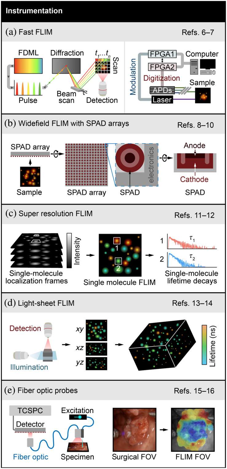
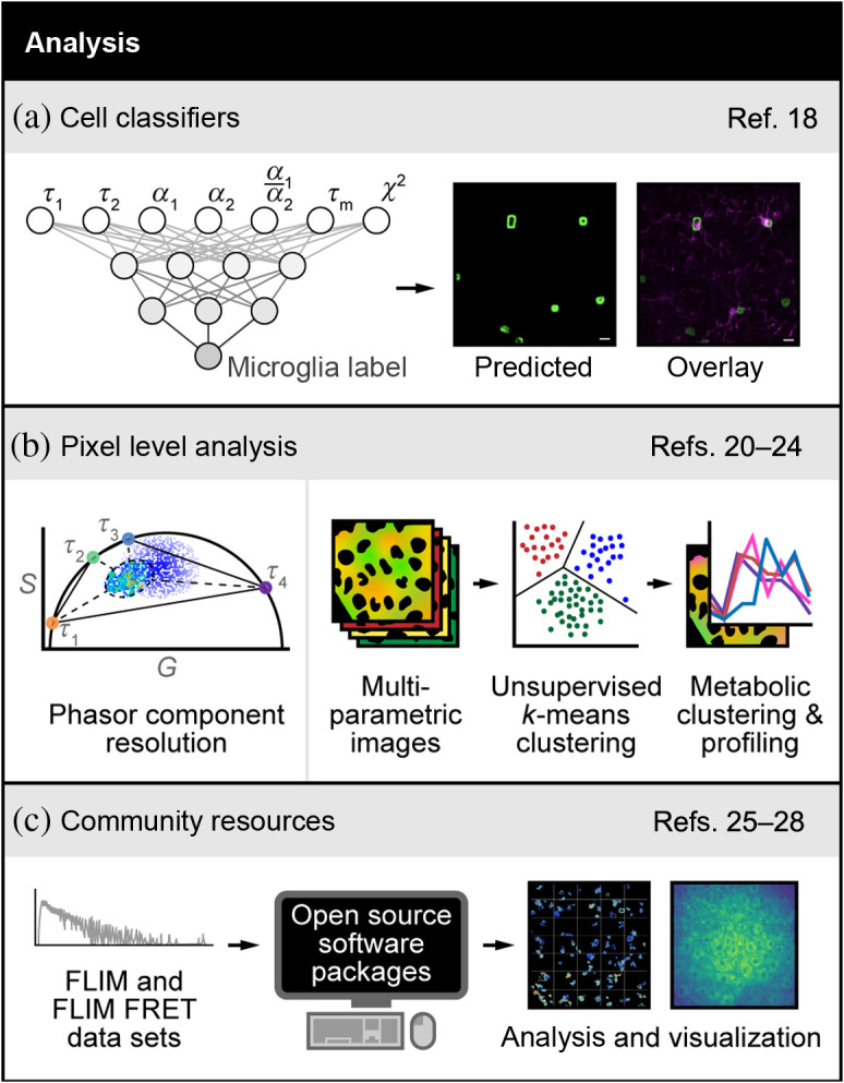
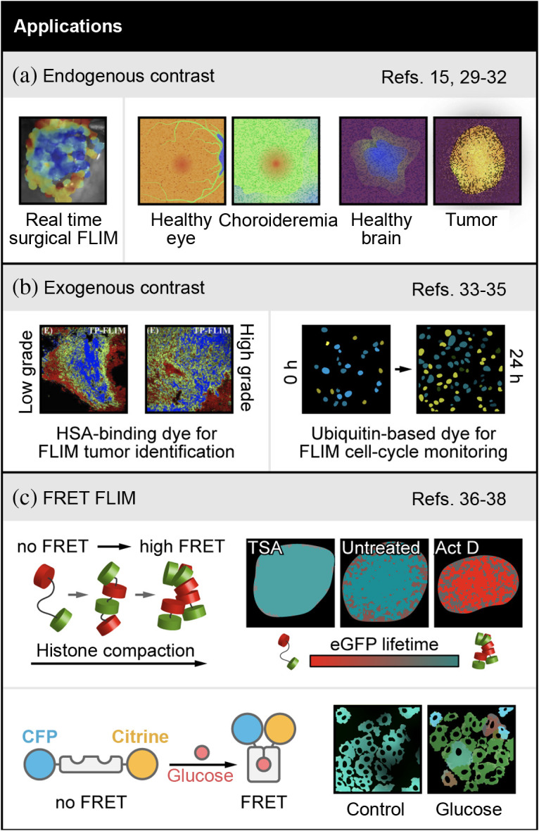
Approach: Innovations in FLIM instrumentation resulted in faster acquisition speeds, rapid imaging over large fields of view, and integration with complementary modalities such as single-molecule microscopy or light-sheet microscopy. There were significant developments in FLIM analysis with machine learning approaches to enhance processing speeds, fit-free techniques to analyze images without a priori knowledge, and open-source analysis resources. The advantages and limitations of these recent instrumentation and analysis techniques are summarized. Finally, applications of FLIM in the last year include label-free imaging in biology, ophthalmology, and intraoperative imaging, FLIM of new fluorescent probes, and lifetime-based Förster resonance energy transfer measurements.

Conclusions: A large number of high-quality publications over the last year signifies the growing interest in FLIM and ensures continued technological improvements and expanding applications in biomedical research.

Keywords: fluorescence lifetime; fluorescence lifetime imaging microscopy; image analysis; microscopy; perspectives.


Figures

Fig. 1
Fig. 1
Advances in FLIM instrumentation. (a) Left panel: simplified schematics of SLIDE microscope that uses swept-source FDML laser that is pulse modulated such that each pulse is encoded both spectrally and in time, resulting in sequential pixel-wise excitation. This technique, along with a fast digitizer, achieves FLIM acquisition at high speed. Adapted with permission from Ref. . Right panel: simplified illustration of frequency domain lifetime imaging system implemented with FPGAs that modulates the excitation laser and digitizes the emissions using fixed gain APDs. Stylized image representing original publication with the permission of AIP Publishing. (b) Schematics of time-gated SPAD array consisting of SPAD photodiode and electronics to measure individual photon arrival times. Cross-sectional view of a SPAD showing key components—diode anode and cathode. FLIM systems implemented with SPAD arrays provide fast FLIM acquisition from large samples. (c) Super-resolved FLIM is acquired with SMLM techniques. Stylized illustration adapted with permission from Ref.  shows intensity frames including single-molecule localizations, single-molecule lifetime map, and histogram of photon arrival times for two indicated single-molecule localizations. (d) Simplified schematic showing FLIM with light-sheet illumination scheme that results in volumetric FLIM, adapted with permission from Ref. . (e) Simplified schematics showing fiber-optic-based FLIM system with TCSPC electronics for time domain FLIM data acquisition. Also shown are white light image of surgical FOV and FLIM FOV with augmented lifetime map overlay. Adapted with permission from Refs.  and . References to all relevant papers for each section have been noted.
Fig. 2
Fig. 2
Advances in FLIM analysis. (a) FLIM of the intrinsically fluorescent metabolic co-factor nicotinamide adenine dinucleotide (phosphate) [NAD(P)H] was combined with ANN-based machine learning to differentiate microglia from other glia cell types in the brain. The two-exponential decay lifetimes (τ1,τ2), weights (α1,α2), mean lifetime (τm=α1τ1+α2τ2), and goodness-of-fit (χ2) were used as input parameters to identify microglia in cell culture mixed with other glial cells and in fixed brain tissue. Adapted with permission from Ref. . (b). Left panel: phasor plots of fluorescence decays provide a visual distribution of fluorescence lifetimes and are derived from a Fourier transformation of the fluorescence lifetime decay with unitless horizontal (G) and vertical (S) axes. Here the phasor method was used to resolve the decay in each pixel of an image in live cells or mouse liver tissues with two or more exponential components without prior knowledge of the values of the components. Adapted with permission from Ref. . Right panel: spectral and lifetime imaging of NAD(P)H and FAD was combined with unsupervised k-means clustering of each pixel to identify regions within cells that have almost uniform metabolic properties. This method detects the cellular mitochondrial turnover and the metabolic activation state of intracellular compartments at the pixel level. Adapted with permission from Ref. . (c) FLIM and FRET decays acquired through any method can be analyzed with open-source software packages to quantify molecular changes and render images. These open-source packages include community forums to educate non-experts and foster continuous development by experts. Images adapted with permission from Refs.  and . References to all relevant papers for each section have been noted.
Fig. 3
Fig. 3
Examples of FLIM in biology and medicine. (a) Left panel: endogenous contrast applications include automated FLIM visualization using FLIMBrush on a clinical head and neck surgery case. Adapted with permission from Ref. . Middle and right panels: FLIM detection of choroideremia (middle) and brain tumors (right) are shown with stylized images that represent changes observed in original publications. (b) Left panel: applications with exogenous contrast include quantitative analysis of HSA concentrations in cryosection from a low-grade and high-grade serous ovarian cancer patient, using the concentration dependent squarine dye (blue = high [HSA], green = mid [HSA], red = low [HSA]). Adapted with permission from Ref. . Right panel: FLIM of an ubiquitination-based dye, FUCCI-Red, which is a lifetime sensor of cell cycle state (blue=S/G2/M, green=G1/S, yellow=G1) shown with stylized images representing changes observed in original publication (c) Top panel: FRET-FLIM applications include a new FLIM-FRET pair sensitive to chromatin compaction. Adapted with permission from Ref. . Pseudocolored chromatin compaction maps of the cells in the top row according to the palette defined below scaled between 2.5 ns (teal, 0% FRET, donor lifetime) and 2 ns (red, 21% FRET, quenched donor). Bottom panel: intracellular glucose concentration imaged with a FLIM-FRET pair. A shift toward red indicates higher intracellular glucose, which can be seen in the glucose condition represented using stylized images. References to all relevant papers for each section have been noted.

References

    1. Datta R., et al. , “Fluorescence lifetime imaging microscopy: fundamentals and advances in instrumentation, analysis, and applications,” J. Biomed. Opt. 25(7), 071203 (2020).JBOPFO10.1117/1.JBO.25.7.071203 - DOI - PMC - PubMed
    1. Gratton E., et al. , “Fluorescence lifetime imaging for the two-photon microscope: time-domain and frequency-domain methods,” J. Biomed. Opt. 8(3), 381–390 (2003).JBOPFO10.1117/1.1586704 - DOI - PubMed
    1. Gratton E., “Measurements of fluorescence decay time by the frequency domain method,” in Perspectives on Fluorescence, pp. 67–80, Springer International Publishing; (2016).
    1. Suhling K., et al. , “Fluorescence lifetime imaging (FLIM): basic concepts and some recent developments,” Med. Photonics 27, 3–40 (2015).10.1016/j.medpho.2014.12.001 - DOI
    1. Suhling K., et al. , “Fluorescence Lifetime Imaging,” in Handbook of Photonics for Biomedical Engineering, pp. 353–405, Springer, Netherlands: (2017).

Publication types

Substances