Review
. 2021 Oct;128(10):e51-e68.
doi: 10.1016/j.ophtha.2021.05.031. Epub 2021 Jul 8.

International Classification of Retinopathy of Prematurity, Third Edition

Affiliations
Review

International Classification of Retinopathy of Prematurity, Third Edition

Michael F Chiang et al. Ophthalmology. 2021 Oct.

Abstract

Purpose: The International Classification of Retinopathy of Prematurity is a consensus statement that creates a standard nomenclature for classification of retinopathy of prematurity (ROP). It was initially published in 1984, expanded in 1987, and revisited in 2005. This article presents a third revision, the International Classification of Retinopathy of Prematurity, Third Edition (ICROP3), which is now required because of challenges such as: (1) concerns about subjectivity in critical elements of disease classification; (2) innovations in ophthalmic imaging; (3) novel pharmacologic therapies (e.g., anti-vascular endothelial growth factor agents) with unique regression and reactivation features after treatment compared with ablative therapies; and (4) recognition that patterns of ROP in some regions of the world do not fit neatly into the current classification system.

Design: Review of evidence-based literature, along with expert consensus opinion.

Participants: International ROP expert committee assembled in March 2019 representing 17 countries and comprising 14 pediatric ophthalmologists and 20 retinal specialists, as well as 12 women and 22 men.

Methods: The committee was initially divided into 3 subcommittees-acute phase, regression or reactivation, and imaging-each of which used iterative videoconferences and an online message board to identify key challenges and approaches. Subsequently, the entire committee used iterative videoconferences, 2 in-person multiday meetings, and an online message board to develop consensus on classification.

Main outcome measures: Consensus statement.

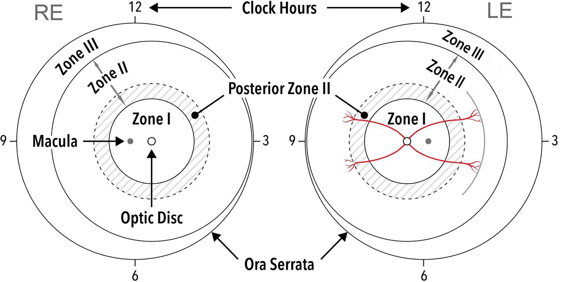
Results: The ICROP3 retains current definitions such as zone (location of disease), stage (appearance of disease at the avascular-vascular junction), and circumferential extent of disease. Major updates in the ICROP3 include refined classification metrics (e.g., posterior zone II, notch, subcategorization of stage 5, and recognition that a continuous spectrum of vascular abnormality exists from normal to plus disease). Updates also include the definition of aggressive ROP to replace aggressive-posterior ROP because of increasing recognition that aggressive disease may occur in larger preterm infants and beyond the posterior retina, particularly in regions of the world with limited resources. ROP regression and reactivation are described in detail, with additional description of long-term sequelae.

Conclusions: These principles may improve the quality and standardization of ROP care worldwide and may provide a foundation to improve research and clinical care.

Keywords: Neonatology; Pediatric ophthalmology; Prematurity; Retina; Retinopathy of prematurity.


Conflict of interest statement

  1. J. Peter Campbell receives research support from Genentech (South San Francisco, CA).

  2. Antonio Capone Jr is an equity owner of Phoenix Technology Group, LLC, a founder and equity owner of Retinal Solutions, LLC and receives research support from AURA Biosciences (Cambridge, MA), Bayer (Leverkusen, Germany), Genentech (South San Francisco, CA), Ionis Pharmaceuticals (Carlsbad, CA), Novartis (Basel, Switzerland) and Regeneron Pharmaceuticals (Tarrytown, NY).

  3. R. V. Paul Chan is on the Scientific Advisory Board for Phoenix Technology (Fremont, CA), a Consultant for Alcon (Ft. Worth, TX), and a Consultant for Novartis (Basel, Switzerland).

  4. Michael F. Chiang was previously a Consultant for Novartis (Basel, Switzerland), an equity owner of InTeleretina (Honolulu, HI), and received research support from Genentech (South San Francisco, CA).

  5. Alistair Fielder is a consultant for Novartis (Basel, Switzerland) and Bayer (Reading, United Kingdom)

  6. Brian Fleck is a consultant for Novartis (Basel, Switzerland).

  7. Mary Elizabeth Hartnett receives research support from the National Institutes of Health (R01EY017011, F01EY01730, EY014800), and is a consultant for Regeneron (Tarrytown, NY).

  8. Domenico Lepore is a Consultant for Novartis (Basel, Switzerland) and Bayer (Leverkusen, Germany).

  9. Şengül Özdek is a consultant for Novartis (Basel, Switzerland), Bayer (Leverkusen, Germany), and Allergan (Dublin, Ireland).

  10. Andreas Stahl receives research support from Novartis (Basel, Switzerland), and is a Consultant for Novartis (Basel, Switzerland) and Bayer (Leverkusen, Germany).

  11. Cynthia A. Toth receives research support from the National Institutes of Health (R01EY025009, U01EY028079, P30EY005722) and from a Research to Prevent Blindness Stein Award, royalties from Alcon (Fort Worth, TX), and is a founding and equity owner of Theia Imaging, LLC.

  12. Wei-Chi Wu is a consultant for Novartis (Basel, Switzerland), Bayer (Leverkusen, Germany), and Allergan (Dublin, Ireland).

Dr. Campbell is supported by research funding from the National Institutes of Health (R01EY19474, K12EY27720), the National Science Foundation (SCH-1622679), and Genentech (South San Francisco, CA). Dr. Capone is an equity owner of Phoenix Technology Group, LLC, a founder and equity owner of Retinal Solutions, LLC and receives research support from AURA Biosciences (Cambridge, MA), Bayer (Leverkusen, Germany), Genentech (South San Francisco, CA), Ionis Pharmaceuticals (Carlsbad, CA), Novartis (Basel, Switzerland) and Regeneron Pharmaceuticals (Tarrytown, NY). Dr. Fleck is a consultant for Novartis (Basel, Switzerland). Dr. Hartnett receives research support from the National Institutes of Health (R01EY017011, F01EY01730, EY014800), and is a consultant for Regeneron (Tarrytown, NY). Dr. Lepore is a Consultant for Novartis (Basel, Switzerland) and Bayer (Leverkusen, Germany). Dr. Özdek is a consultant for Novartis (Basel, Switzerland), Bayer (Leverkusen, Germany), and Allergan (Dublin, Ireland). Dr. Stahl receives research support from Novartis (Basel, Switzerland), and is a Consultant for Novartis (Basel, Switzerland) and Bayer (Leverkusen, Germany). Dr. Toth receives research support from the National Institutes of Health (R01EY025009, U01EY028079, P30EY005722) and from a Research to Prevent Blindness Stein Award, royalties from Alcon (Fort Worth, TX), and is a founding and equity owner of Theia Imaging, LLC. Dr. Wu is a consultant for Novartis (Basel, Switzerland), Bayer (Leverkusen, Germany), and Allergan (Dublin, Ireland).

Figures

Figure 1.
Figure 1.. Schema of right eye (RE) and left eye (LE) showing zone borders and clock hour sectors used to describe the location of vascularization and extent of retinopathy.
Solid circles represent borders of zones I-III, and dotted circles represent borders of posterior zone II (two disc diameters beyond zone I). A hypothetical example of examination findings is shown in the left eye, representing approximately three clock hours of stage 1 disease in zone II (note single line on drawing to document presence of stage 1 disease).
Figure 2.
Figure 2.. Wide-angle fundus photographs demonstrating examples of plus disease and pre-plus disease. Note varying levels of vascular abnormality, which are assessed in the central retina within the region of zone I.
(A) Mild pre-plus disease, with more arterial tortuosity and venous dilation than normal. (B) Pre-plus disease, with notable arterial tortuosity but minimal venous dilation. (C) Pre-plus disease, with moderate arterial tortuosity and venous dilation, but considered by majority of committee members to be insufficient for plus disease. (D) Plus disease with notable venous dilation and arterial tortuosity. Note that plus disease is out of proportion to visible peripheral findings, suggestive of flat neovascularization (stage 3, white arrows). (E) Severe plus disease, with dilation and tortuosity of both arteries and veins. (F) Severe plus disease. Note presence of ill-defined posterior flat stage 3 (arrows), which combined with severe plus disease is typical of aggressive ROP (A-ROP).
Figure 3.
Figure 3.. Continuous spectrum of vascular severity in ROP from normal to plus disease.
All 34 members of this committee graded 30 images as normal, pre-plus, or plus. Experts’ opinions varied as to the level of disease severity that constitutes pre-plus and plus disease. (A) Six representative images are displayed, where color scale on top reflects the average grading of committee members (from green = normal to red = plus), and demonstrates that vascular severity presents on a continuum. (B) Nine representative segmented images are displayed, where color scale represents mean vascular severity grading by committee members for each image (from green = normal to red = plus), and demonstrates that vascular severity presents on a continuum.
Figure 4.
Figure 4.. Wide-angle fundus photograph demonstrating incomplete vascularization into zone II in the right eye of a premature infant at risk for retinopathy of prematurity.
Note progressive tapering and termination of retinal vascular arcades (white arrows).
Figure 5.
Figure 5.. Wide-angle fundus photographs demonstrating examples of acute retinopathy of prematurity (ROP) stages 1–3.
(A) Stage 1 demarcation line at border between vascular and avascular retina (white arrows). (B) Stage 1 demarcation line (white arrows) and associated notch (black arrowheads) between vascular arcades which would be considered zone I secondary to notch. Note pre-plus disease with mild retinal vascular tortuosity and dilation. (C) Stage 2 ridge, which is raised (white arrows) and thicker than stage 1. (D) Stage 2 ridge. Note “popcorn” lesions posterior to ridge (arrows) and pre-plus disease with mild vascular tortuosity and dilation. (E) Stage 3 disease with extraretinal neovascularization (white arrows). Note plus disease with vascular tortuosity and dilation. (F) Eye with both stage 2 (black arrowheads) and stage 3 (white arrowheads), and associated “popcorn” (white arrows). Note plus disease with vascular tortuosity and dilation.
Figure 6.
Figure 6.. Optical coherence tomography (OCT) images demonstrating examples of retinopathy of prematurity stages 1–3.
OCT cross-sectional B-scans (left side) are extracted from the raster scans that make up the OCT volume, at the location of the green-line (right side). The en face OCT images (right side) are summed from the OCT volume, and blood in retinal vessels casts a shadow across the underlying retina and choroid. (A) Stage 1 demarcation line (white arrows) on cross-sectional B-scan OCT image (left side) align with the retinal vascular-avascular junction (right side, note the vascular retina is to the left in all images). (B) Stage 2 ridge (white arrows) on cross-sectional B-scan OCT image (left side) at a site of focal thickening and bulge of inner retinal layers, aligns with the en face (right side) wider dark border of the vascular-avascular junction. (C) Stage 3 extraretinal neovascular proliferation at (white arrows) and posterior to (arrowheads) the vascular-avascular junction on cross-sectional B-scan OCT image (left side) and on en face view (right side). Note that blood in the extraretinal vessels also casts a shadow across the underlying retina and choroid.
Figure 7.
Figure 7.. Aggressive retinopathy of prematurity (A-ROP).
(A) A-ROP with severe vasoconstriction, capillary nonperfusion, non-physiologic dilated vascular loops and arterio-venous shunts, and plus disease in zone I. (B) A-ROP with border between vascular and avascular retina in zone I, dilated vascular loops (white arrows), diffuse flat extraretinal neovascularization most prominent superotemporally, and severe plus disease. Note absence of a typical stage 3 lesion. (C) A-ROP in zone I with severe plus disease, flat extraretinal neovascularization with fibrosis and early contraction superiorly (white arrowheads), and intraretinal and vitreous hemorrhage superotemporally. (D) and (E) are two examples of wide-angle fundus photographs (left sides) demonstrating A-ROP with ill-defined junction between vascular and avascular retina in zone I (white arrows), and severe plus disease. Fluorescein angiography (right sides) demonstrate significant vaso-obliteration with capillary nonperfusion. Note that there are no typical ROP lesion appearances and that there are vaso-attenuated areas posterior to the ridge.
Figure 8.
Figure 8.. Wide-angle fundus photographs demonstrating examples of retinopathy of prematurity stage 4.
(A) Stage 4A in temporal retina. Traction on extraretinal neovascularization leads to retinal elevation (white dots), which may be recognized during ophthalmoscopy by subtle change in brightness and loss of visible retinal pigment epithelium granularity and choriocapillaris detail. Note that approximate foveal center (asterisk) is not elevated, and that the extraretinal neovascularization (white arrows) may be significantly more peripheral than posterior extent of the detachment. (B) Stage 4A with 360-degree tractional retinal detachment in area of the peripheral ridge. (C) Stage 4B detachment involving the macula. Note straightening of the arcuate vessels and dragged optic disc appearance. (D) Stage 4B detachment with associated subretinal hemorrhage and lipid exudation into the macula. (E) Volcano shaped stage 4B. In eyes with posterior ROP, contraction of pathologic neovascularization can result in detachment of vascularized retina into a volcano-shaped configuration.
Figure 9.
Figure 9.. Optical coherence tomography (OCT) images demonstrating examples of stage 4 disease.
OCT cross-sectional B-scans (left side) are extracted from the raster of scans that make up the OCT volumes, viewed as summed en face (middle) with corresponding color photographs (right side). Note that in the color photographs, it is difficult to discern the extent of retinal detachment, especially foveal involvement. In en face OCT and color photos, the location of the fovea (asterisk) and margin of detachment (white dots) are determined from OCT B-scans. Blood in retinal vessels casts a shadow across the underlying retina and choroid in the en face views. (A) Stage 4A retinal detachment. On the en face OCT image (middle), yellow line demonstrates location of B-scan superior to the foveal center (upper left side, yellow box), green line demonstrates location of B-scan through the foveal center (bottom left side, green box). Note the attached retina on B-scan at the foveal center (asterisk), along with intraretinal exudates (hyperreflective dots), peripheral retinal detachment, and retinoschisis. (B) Stage 4B retinal detachment. Note detachment of retina at the foveal center (asterisks), which was very difficult to appreciate on ophthalmoscopic examination.
Figure 10.
Figure 10.. Images demonstrating examples of stage 5 retinopathy of prematurity.
(A) Wide-angle fundus photograph showing stage 5A, characterized by a total retinal detachment with visible optic disc. Note open-funnel configuration. (B) Wide-angle fundus photograph showing Stage 5B, with no view of optic disc because of fibrovascular tissue. (C) External photograph of the normal anterior segment in stage 5B (left side), with no view of optic disc or retina secondary to retrolental fibrovascular tissue. B-scan ultrasonography (right side) reveals total retinal detachment with a posteriorly closed funnel configuration. (D) External photograph showing anterior segment characteristic of Stage 5C with anterior lens displacement, marked anterior chamber shallowing, central irido-capsule-endothelial adhesion, and central corneal opacification (asterisk) that prevent view of closed-funnel retinal detachment.
Figure 11.
Figure 11.. Wide-angle fundus photographs demonstrating examples of ROP regression.
(A) Regression after laser treatment. Image on left is pre-treatment, showing stage 3 with plus disease. Image in middle is 1 week post-treatment, showing that stage 3 is thinner and whiter. Image on right is 1 month post-treatment, showing disease regression. (B) Regression of plus disease after anti-VEGF injection for aggressive ROP (A-ROP). Image on left is pre-treatment, showing plus disease and flat neovascularization (stage 3). Image on right is 2 weeks post-treatment, showing improvement in plus disease with no visible ROP lesion. (C) Regression after anti-VEGF injection. Image on left is pre-treatment. Image on right is 4 weeks post-treatment, showing absence of stage 3 and improvement in plus disease, with vascularization into peripheral avascular retina. Note the circumferential anastomosis in the area of original stage 3, along with reactivated stage 1 more anteriorly.
Figure 12.
Figure 12.. Examples of persistent avascular retina (PAR) and long-term sequelae of ROP.
(A) Combined tractional and exudative detachment in an 18 year-old with a history of untreated ROP whose fellow eye was phthisical from ROP. (B) Ultra-widefield fluorescein angiogram (UWFFA) demonstrating PAR (asterisks) in a 7 year-old child with a history of spontaneously regressed ROP. Note the abnormal vascular configuration, particularly inferotemporally (circle). (C) UWFFA from a 7 year-old with spontaneously regressed ROP but with PAR and leakage in incompletely regressed stage 3 inferotemporally (asterisks). (D) Ultra-widefield fundus image (left side) displays an incompletely regressed ridge (white arrowheads) with PAR (asterisks) in a 15 year-old child with a history of extreme prematurity and no prior ROP treatment. Two years later (right side), the patient presented with a macula-involving rhegmatogenous detachment. The fellow eye had a similar appearance and disease course. (E) Optical coherence tomography angiography (OCTA) image of an incompletely developed foveal contour (left) and poorly-defined foveal avascular zone (right) in a 7 year-old with history of type 1 ROP treated with laser.
Figure 13.
Figure 13.. Examples of ROP reactivation.
(A) Image at 38 weeks post-menstrual age (PMA) after intravitreal anti-vascular endothelial growth factor (anti-VEGF) injection at 32 weeks PMA with vascularization into peripheral avascular retina. Demarcation line (arrow) at the leading edge is reactivated stage 1. (B) Image of left eye at 100 weeks PMA after treatment with intravitreal anti-VEGF injection at 38 weeks PMA. There was vascularization into the peripheral avascular retina. There are often notable vascular abnormalities at the site of the original ridge, and in some cases residual fibrosis (asterisk), which is not indicative of reactivation unless accompanied by increasing vascular activity. (C) Image of vascularization into peripheral avascular retina with reactivated stage 1 disease (arrow) at 68 weeks PMA, after treatment with intravitreal anti-VEGF injection at 37 weeks PMA. Note multiple circumferential vascular loops at the site of the original ridge (asterisk). (D) Reactivation in a right eye at 67 weeks PMA, which had undergone intravitreal anti-VEGF injection at 33 weeks and again at 52 weeks PMA. There is reactivated stage 3 (asterisk) posterior to the leading edge of vascularization (arrow). (E) Left eye with reactivated stage 3 at the leading edge (arrow) at 50 weeks PMA, after intravitreal anti-VEGF injection at 36 weeks PMA. Vascularization into peripheral avascular retina has occurred between the original ridge (asterisks) and anterior reactivation. (F) Fluorescein angiogram at 45 weeks PMA of a left eye that had intravitreal anti-VEGF injection at 34 weeks PMA. There is leakage both at sites of leading edge reactivation (arrow), and at the original border (asterisk). (G) Right eye with zone I disease treated with intravitreal anti-VEGF injection at 34 weeks PMA (left side, arrow), and which appeared regressed on clinical exam at 38 weeks PMA (middle image, arrow). At 51 weeks PMA, the eye developed reactivated stage 3 at the same site (right side, arrow), without evidence of vascularization into peripheral avascular retina.

Comment in

References

    1. Reese AB, King MJ, Owens WC. Classification of retrolental fibroplasia. Am J Ophthalmol. 1953;36(10):1333–1335. - PubMed
    1. The Committee for the Classification of Retinopathy of Prematurity. An international classification of retinopathy of prematurity. Arch Ophthalmol. 1984;102(8):1130–1134. - PubMed
    1. Cryotherapy for Retinopathy of Prematurity Cooperative Group. Multicenter trial of cryotherapy for retinopathy of prematurity. Preliminary results. Arch Ophthalmol. 1988;106(4):471–479. - PubMed
    1. ICROP Committee for classification of late stages of ROP: An international classification of retinopathy of prematurity: II The classification of retinal detachment. Arch Ophthalmol. 1987;105:906–912. - PubMed
    1. Committee for the Classification of Retinopathy of Prematurity. The International Classification of Retinopathy of Prematurity Revisited. Arch Ophthal. 2005;123(7):991. - PubMed

Publication types