

Neuroinflammation-Driven Lymphangiogenesis in CNS Diseases
- PMID: 34248503
- PMCID: PMC8261156
- DOI: 10.3389/fncel.2021.683676
Neuroinflammation-Driven Lymphangiogenesis in CNS Diseases
Abstract
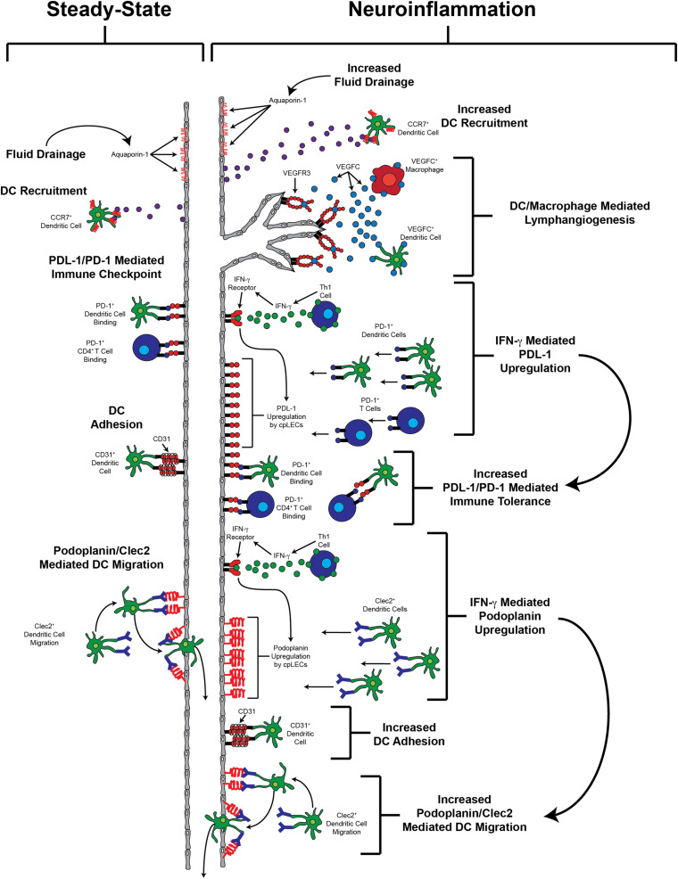
The central nervous system (CNS) undergoes immunosurveillance despite the lack of conventional antigen presenting cells and lymphatic vessels in the CNS parenchyma. Additionally, the CNS is bathed in a cerebrospinal fluid (CSF). CSF is continuously produced, and consequently must continuously clear to maintain fluid homeostasis despite the lack of conventional lymphatics. During neuroinflammation, there is often an accumulation of fluid, antigens, and immune cells to affected areas of the brain parenchyma. Failure to effectively drain these factors may result in edema, prolonged immune response, and adverse clinical outcome as observed in conditions including traumatic brain injury, ischemic and hypoxic brain injury, CNS infection, multiple sclerosis (MS), and brain cancer. Consequently, there has been renewed interest surrounding the expansion of lymphatic vessels adjacent to the CNS which are now thought to be central in regulating the drainage of fluid, cells, and waste out of the CNS. These lymphatic vessels, found at the cribriform plate, dorsal dural meninges, base of the brain, and around the spinal cord have each been implicated to have important roles in various CNS diseases. In this review, we discuss the contribution of meningeal lymphatics to these processes during both steady-state conditions and neuroinflammation, as well as discuss some of the many still unknown aspects regarding the role of meningeal lymphatics in neuroinflammation. Specifically, we focus on the observed phenomenon of lymphangiogenesis by a subset of meningeal lymphatics near the cribriform plate during neuroinflammation, and discuss their potential roles in immunosurveillance, fluid clearance, and access to the CSF and CNS compartments. We propose that manipulating CNS lymphatics may be a new therapeutic way to treat CNS infections, stroke, and autoimmunity.
Keywords: CNS; CNS autoimmunity; CNS infection; CNS trauma; cribriform plate; lymphoid vessels; meningeal lymphatics.
Copyright © 2021 Hsu, Laaker, Sandor and Fabry.
Conflict of interest statement
The authors declare that the research was conducted in the absence of any commercial or financial relationships that could be construed as a potential conflict of interest.
Figures
References
Publication types
Grants and funding
LinkOut - more resources
Full Text Sources