. 2021 Jun 23:15:696376.
doi: 10.3389/fnana.2021.696376. eCollection 2021.

Ultrastructural Characterization of Human Oligodendrocytes and Their Progenitor Cells by Pre-embedding Immunogold

Affiliations

Ultrastructural Characterization of Human Oligodendrocytes and Their Progenitor Cells by Pre-embedding Immunogold

María J Ulloa-Navas et al. Front Neuroanat. .

Abstract

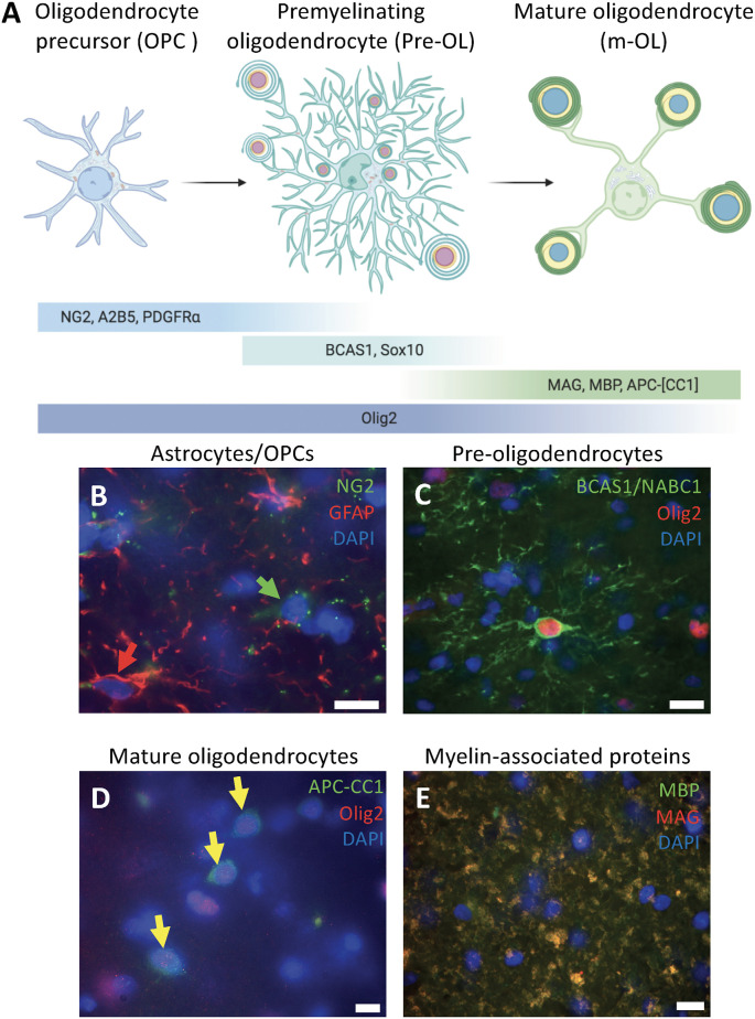
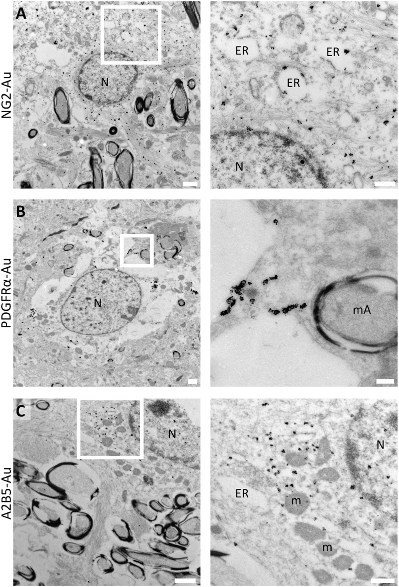
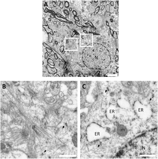
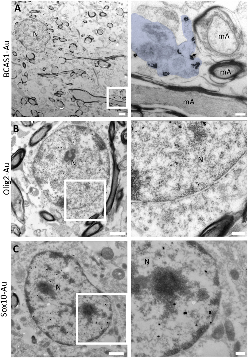
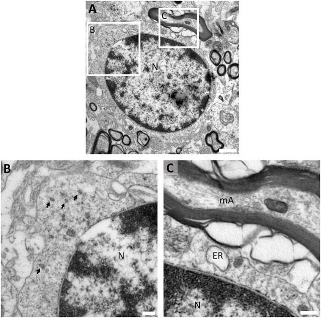
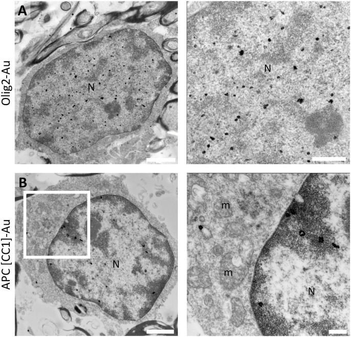
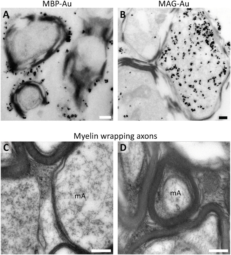
Oligodendrocytes are the myelinating cells of the central nervous system. They provide trophic, metabolic, and structural support to neurons. In several pathologies such as multiple sclerosis (MS), these cells are severely affected and fail to remyelinate, thereby leading to neuronal death. The gold standard for studying remyelination is the g-ratio, which is measured by means of transmission electron microscopy (TEM). Therefore, studying the fine structure of the oligodendrocyte population in the human brain at different stages through TEM is a key feature in this field of study. Here we study the ultrastructure of oligodendrocytes, its progenitors, and myelin in 10 samples of human white matter using nine different markers of the oligodendrocyte lineage (NG2, PDGFRα, A2B5, Sox10, Olig2, BCAS1, APC-(CC1), MAG, and MBP). Our findings show that human oligodendrocytes constitute a very heterogeneous population within the human white matter and that its stages of differentiation present characteristic features that can be used to identify them by TEM. This study sheds light on how these cells interact with other cells within the human brain and clarify their fine characteristics from other glial cell types.

Keywords: BCAS1; OPCs; human oligodendrocytes; immunogold; oligodendrocytes; transmission electron microscopy.


Conflict of interest statement

The authors declare that the research was conducted in the absence of any commercial or financial relationships that could be construed as a potential conflict of interest.

Figures

Figure 1
Figure 1
Differentiation stages of oligodendrocytes in human white matter. (A) Schematic diagram of the oligodendrocyte differentiation process representing the morphology, organelle distribution, and markers to identify each of the three stages: oligodendrocyte precursor cells (OPCs), premyelinating oligodendrocytes (pre-OLs), and mature oligodendrocytes. (B) Human white matter labeled for the astrocytic marker GFAP (red arrow) and the OPC marker NG2 (green arrow) showing that these are two different populations within the white matter of a 39-year-old male (PB4). (C) Human white matter labeled for pre-myelinating oligodendrocyte markers BCAS1/NABC1 and Olig2 co-expressed in highly ramified cells from a 5-year-old female (PB19). (D) Mature oligodendrocytes labeled for Olig2 and APC-(CC1; yellow arrows) from a white matter sample of a 5-year-old female (PB19). (E) Myelin wrapped axons co-expressing MAG and MBP from a white matter sample of a 39-year-old male (PB4). Scale bars: 50 μm.
Figure 2
Figure 2
Immunoelectron microscopy for human OPC molecular markers. (A) NG2 label in large electron-lucent cells in the white matter. Inset shows NG2 subcellular distribution in cytoplasmic intermediate filaments and in short dilated endoplasmic reticulum cisternae in a sample of a 2-year-old male sample (PB20). (B) PDGFRα is expressed in discrete clusters in the plasma membrane of OPCs in the white matter of a 6-year-old male (PB3). (C) OPCs in the white matter labeled with A2B5. Immunogold shows the subcellular localization of this marker in the cytoplasmic portion of the cells and is also associated with the endoplasmic reticulum in the white matter of a 5-year-old female (PB19). N, Nucleus; ER, endoplasmic reticulum; mA, myelinated axon; m, mitochondria. Scale bars: panoramic micrographs, 1 μm; insets: 250 nm.
Figure 3
Figure 3
Ultrastructural characterization of OPCs in the human white matter. (A) Panoramic micrograph of an OPC showing a large electron-lucent star-shaped cell with electron-lucent nucleus (with abundant euchromatin) intermingled between myelinated axons in the white matter. (B) OPCs present abundant filamentous processes, and (C) an electron-lucent cytoplasm with abundant short dilated endoplasmic reticulum cisternae, mitochondria and ribosomes. The micrograph proceeds from the white matter of a 27-year-old male (PB8) fixed with 2% PFA-2.5% Glutaraldehyde. N, Nucleus; ER, endoplasmic reticulum; m, mitochondria; IF, intermediate filaments; arrows, polyribosomes. Scale bars: (A), 2 μm; (B,C), 500 nm.
Figure 4
Figure 4
Pre-embedding immunogold labeling for pre-myelinating oligodendrocytes. (A) BCAS1/NABC1 labeled pre-myelinating oligodendrocyte. BCAS1 is observed in the plasma membrane of the cell and in processes in direct contact with thin myelin sheets (blue pseudocolor) in the white matter of a 2-year-old female (PB9). (B) Olig2 labels the uncondensed chromatin of electron-lucent cells with scant cytoplasm in the white matter of a 32-year-old male (PB16). (C) The transcription factor SOX10 labels the nucleus of pre-myelinating oligodendrocytes in the white matter of a 13-year-old female (PB11). N, Nucleus; mA, myelinated axon. Scale bars: panoramic micrographs: 1 μm; insets: 250 nm.
Figure 5
Figure 5
Fine structure of human pre-myelinating oligodendrocytes. (A) Pre-myelinating oligodendrocytes are highly ramified cells with scant electron-lucent cytoplasm and slightly condensed chromatin. (B) Their cytoplasm contains abundant polyribosomes, (C) a few small mitochondria and short dilated endoplasmic reticulum. These cells are in direct contact with myelin sheets The micrograph proceeds from the white matter of a 9-year-old male (PB2) fixed with 2% PFA–2.5% Glutaraldehyde. N, Nucleus; ER, endoplasmic reticulum; arrows, polyribosomes. T Scale bars: (A), 1 μm; (B,C), 250 nm.
Figure 6
Figure 6
Fully differentiated oligodendrocytes display Olig2 and APC-(CC1) labeling. (A) Olig2 expression in mature oligodendrocytes is restricted to the uncondensed chromatin within the nucleus in the white matter of a 44-year-old male (PB12). (B) The RNA-binding protein quaking7 labeled with APC-(CC1) was found in the condensed chromatin portion as well as in non-condensed chromatin; faint labeling was also observed in the plasma membrane in the white matter of a 13-year-old female (PB11). N, Nucleus; m, mitochondria. Scale bars: panoramic micrographs, 1 μm; insets: 250 nm.
Figure 7
Figure 7
Mature oligodendrocytes are electron-dense cells in the human white matter. (A) Mature oligodendrocytes are small electron-dense cells with a broad distribution in the white matter. These cells are intermingled between myelin-wrapped axons. (B) Chromatin condensation occurs bound to the inner nuclear membrane in mature oligodendrocytes. (C) Cytoplasmic organization in this population displays characteristic short dilated endoplasmic reticulum, few mitochondria, dense degradation vesicles, and polyribosomes. The micrograph proceeds from the white matter of a 6-year-old male (PB3) fixed with 2% PFA-2.5% Glutaraldehyde. N, Nucleus; ER, endoplasmic reticulum. Scale bars: (A), 1 μm; (B–C), 250 nm.
Figure 8
Figure 8
Ultrastructural analysis of myelin in the human pre-mortem white matter. (A) Pre-embedding immunogold labeling shows that MBP is located in the outermost layer of the myelinated axon in the cerebral white matter of a 5-year-old female (PB19). (B) MAG is observed in the internal layer of the myelinated axon in direct contact with the axon in the white matter of a 5-year-old female (PB19). (C) Glutaraldehyde fixation allows a better analysis of the fine structure of myelin sheets in different stages of the myelination process, allowing to discern different myelin thickness. (D) Furthermore, it allows the visualization of the number of myelin layers. Ultrastructural analysis reveals that myelin is tightly packed, and exhibits variable periaxonal space. (C,D) Micrographs proceed from the white matter of a 2-year-old female (PB9) fixed with 2% PFA-2.5% Glutaraldehyde. mA, myelinated axon. Scale bars: 250 nm.

Similar articles

Cited by

References

    1. Amaral A. I., Tavares J. M., Sonnewald U., Kotter M. R. N. (2016). Oligodendrocytes: development, physiology and glucose metabolism. Adv. Neurobiol. 13, 275–294. 10.1007/978-3-319-45096-4_10 - DOI - PubMed
    1. Barateiro A., Fernandes A. (2014). Temporal oligodendrocyte lineage progression: In vitro models of proliferation, differentiation and myelination. Biochim. Biophys. Acta 1843, 1917–1929. 10.1016/j.bbamcr.2014.04.018 - DOI - PubMed
    1. Berg G. J., Schachner M. (1982). Electron-microscopic localization of A2B5 cell surface antigen in monolayer cultures of murine cerebellum and retina. Cell Tissue Res. 224, 637–645. 10.1007/BF00213758 - DOI - PubMed
    1. Bribián A., Medina-Rodríguez E. M., Josa-Prado F., García-Álvarez I., Machín-Díaz I., Esteban P. F., et al. . (2020). Functional heterogeneity of mouse and human brain OPCs: relevance for preclinical studies in multiple sclerosis. J. Clin. Med. Res. 9:1681. 10.3390/jcm9061681 - DOI - PMC - PubMed
    1. Bin J. M., Harris S. N., Kennedy T. E. (2016). The oligodendrocyte-specific antibody “CC1” binds quaking 7. J. Neurochem. 139, 181–186. 10.1111/jnc.13745 - DOI - PubMed