Review
. 2021 Jun 23:15:703835.
doi: 10.3389/fnana.2021.703835. eCollection 2021.

The Enigmatic Reissner's Fiber and the Origin of Chordates

Affiliations
Review

The Enigmatic Reissner's Fiber and the Origin of Chordates

Francisco Aboitiz et al. Front Neuroanat. .

Abstract

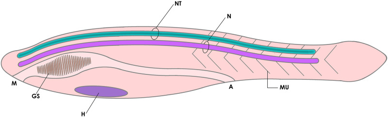
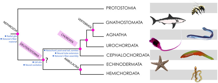
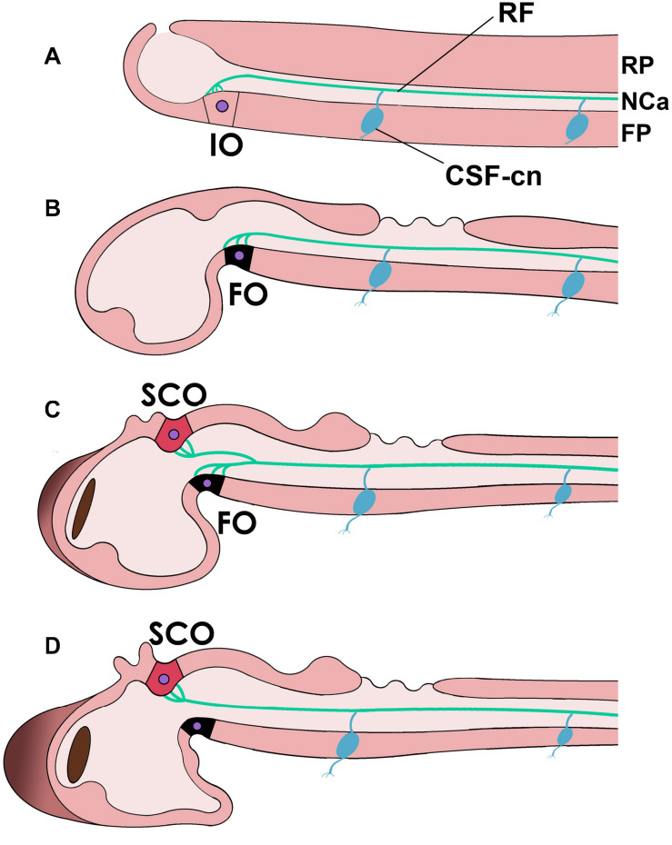
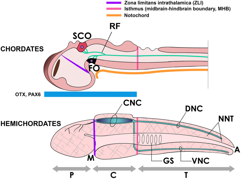
Reissner's fiber (RF) is a secreted filament that floats in the neural canal of chordates. Since its discovery in 1860, there has been no agreement on its primary function, and its strong conservation across chordate species has remained a mystery for comparative neuroanatomists. Several findings, including the chemical composition and the phylogenetic history of RF, clinical observations associating RF with the development of the neural canal, and more recent studies suggesting that RF is needed to develop a straight vertebral column, may shed light on the functions of this structure across chordates. In this article, we will briefly review the evidence mentioned above to suggest a role of RF in the origin of fundamental innovations of the chordate body plan, especially the elongation of the neural tube and maintenance of the body axis. We will also mention the relevance of RF for medical conditions like hydrocephalus, scoliosis of the vertebral spine and possibly regeneration of the spinal cord.

Keywords: Reissner’s fiber; cerebrospinal fluid; chordates; neural tube; notochord; swimming behavior; vertebrates.


Conflict of interest statement

The authors declare that the research was conducted in the absence of any commercial or financial relationships that could be construed as a potential conflict of interest.

Figures

FIGURE 1
FIGURE 1
Diagram of the basic elements of the chordate body plan, featuring a tadpole-like animal with a fibrous notochord (N) and a hollow neural tube (NT) in the dorsal side (cavitation is shown in dark green), a muscular tail (MU) for swimming, and gill slits (GS, a character shared with other deuterostomes). A, anus; H, heart; M, mouth.
FIGURE 2
FIGURE 2
Phylogeny of deuterostomes, indicating the points of origin of the characters discussed in this article. Most phylogenetic analyses place Deuterostomia as a monophyletic group (as depicted here), with Chordata and ambulacria as sister groups. However, some recent findings using large scale genetic databases claim that there is no evidence for Deuterostomia as a monophyletic group (Kapli et al., 2021). Hence the interrogation mark shown at this node. The main conclusions of this article are consistent with both views.
FIGURE 3
FIGURE 3
Reissner’s fiber (RF, green) in different chordates and during development. (A) In the neural tube of cephalochordates RF is secreted by the infundibular organ of the floor plate (FP) (IO) and floats in the CSF-filled neural or central canal (NCa). (B–D) Reissner’s fiber in teleost fish [(B), early embryonic; (C), middle stage; (D), advanced]. Note that in teleosts, the flexural organ (FO, possibly homologous to the IO in cephalochordates) is replaced by the subcommissural organ (SCO) in the roof plate (RP) as the secreting organ of RF. Note also the CSF-contacting neurons (CSF-cn) with an axon (inferior process) and a dendrite or cilium that enters the CSF and contacts RF.
FIGURE 4
FIGURE 4
Above, the neural tube of a chordate (embryonic fish) showing the subcommissural organ (SCO) and the flexural organ (FO), sites of origin of RF. These organs are located in the mesencephalic-diencephalic boundary, which is included in the embryonic region bound by the zona limitans intrathalamica (ZLI) anteriorly and the midbrain-hindbrain boundary (MHB) posteriorly. This region is strongly Otx-positive in early development and for this reason has been considered homologous to the location of the collar region (C) of the hemichordate body (below), displaying the same morphogenetic boundaries (ZLI and MHB homologs) and containing a hollow collar nerve cord (CNC) (Pani et al., 2012; Lowe et al., 2015). Beside the CNC, hemichordates display dorsal and ventral nerve cords (DNC, VNC) in the body trunk (T) and a diffuse neural network (NNT, hatched areas) covering the body (Holland et al., 2015). A, anus; GS, gill slits; M, mouth; P, proboscis.

References

    1. Aboitiz F., Montiel J. (2007). Origin and evolution of the vertebrate telencephalon, with special reference to the mammalian neocortex. Adv. Anat. Embryol. Cell Biol. 193 1–112. 10.1007/978-3-540-49761-5 - DOI - PubMed
    1. Alibardi L. (2021). Tail regeneration in Lepidosauria as an exception to the generalized lack of organ regeneration in amniotes. Exp. Zool. B. Mol. Dev. Evol. 336 145–164. 10.1002/jez.b.22901 - DOI - PubMed
    1. Arendt D., Benito-Gutierrez E., Brunet T., Marlow H. (2015). Gastric pouches and the mucociliary sole: setting the stage for nervous system evolution. Philos. Trans. R. Soc. Lond. B Biol. Sci. 370:20150286. 10.1098/rstb.2015.0286 - DOI - PMC - PubMed
    1. Bearce E. A., Grimes D. T. (2021). On being the right shape: roles for motile cilia and cerebrospinal fluid flow in body and spine morphology. Semin. Cell Dev. Biol. 110 104–112. 10.1016/j.semcdb.2020.07.005 - DOI - PubMed
    1. Beaster-Jones L., Kaltenbach S. L., Koop D., Yuan S., Chastain R., Holland L. Z. (2008). Expression of somite segmentation genes in amphioxus: a clock without a wavefront? Dev. Genes Evol. 218 599–611. 10.1007/s00427-008-0257-5 - DOI - PubMed

LinkOut - more resources