

The Cerebellum Is Related to Cognitive Dysfunction in White Matter Hyperintensities
- PMID: 34248601
- PMCID: PMC8261068
- DOI: 10.3389/fnagi.2021.670463
The Cerebellum Is Related to Cognitive Dysfunction in White Matter Hyperintensities
Abstract
Objective: White matter hyperintensities (WMHs) on magnetic resonance imaging (MRI) is frequently presumed to be secondary to cerebral small vessel disease (CSVD) and associated with cognitive decline. The cerebellum plays a key role in cognition and has dense connections with other brain regions. Thus, the aim of this study was to investigate if cerebellar abnormalities could occur in CSVD patients with WMHs and the possible association with cognitive performances.
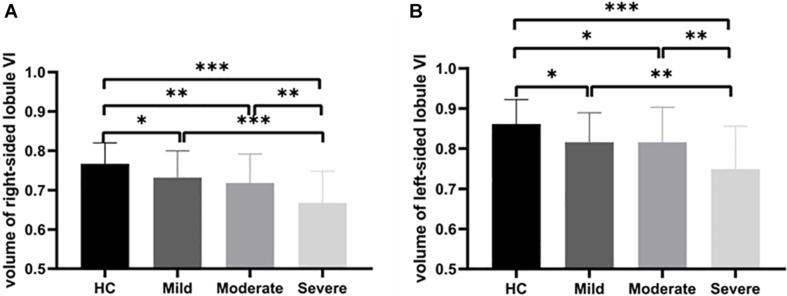
Methods: A total of 104 right-handed patients with WMHs were divided into the mild WMHs group (n = 39), moderate WMHs group (n = 37), and severe WMHs group (n = 28) according to the Fazekas scale, and 36 healthy controls were matched for sex ratio, age, education years, and acquired resting-state functional MRI. Analysis of voxel-based morphometry of gray matter volume (GMV) and seed-to-whole-brain functional connectivity (FC) was performed from the perspective of the cerebellum, and their correlations with neuropsychological variables were explored.
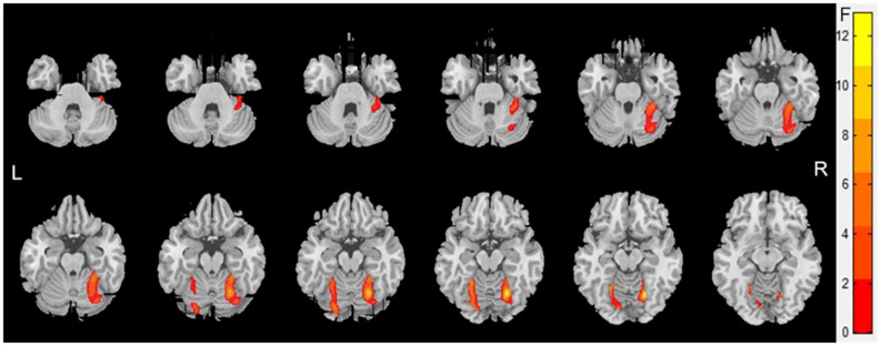
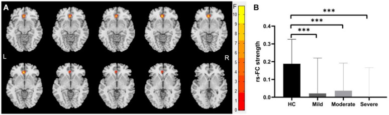
Results: The analysis revealed a lower GMV in the bilateral cerebellum lobule VI and decreased FC between the left- and right-sided cerebellar lobule VI with the left anterior cingulate gyri in CSVD patients with WMHs. Both changes in structure and function were correlated with cognitive impairment in patients with WMHs.
Conclusion: Our study revealed damaged GMV and FC in the cerebellum associated with cognitive impairment. This indicates that the cerebellum may play a key role in the modulation of cognitive function in CSVD patients with WMHs.
Keywords: cerebellum; magnetic resonance imaging; resting-state functional connectivity; voxel-based morphometry; white matter hyperintensities.
Copyright © 2021 Cao, Nie, Zhang, Chen, Wang, Liu, Mo, Du, Hu, Tian, Wei and Wang.
Conflict of interest statement
The authors declare that the research was conducted in the absence of any commercial or financial relationships that could be construed as a potential conflict of interest.
Figures
Similar articles
-
Altered Functional Connectivity Patterns of Parietal Subregions Contribute to Cognitive Dysfunction in Patients with White Matter Hyperintensities.J Alzheimers Dis. 2021;84(2):659-669. doi: 10.3233/JAD-210315. J Alzheimers Dis. 2021. PMID: 34569947
-
Abnormal Degree Centrality in White Matter Hyperintensities: A Resting-State Functional Magnetic Resonance Imaging Study.Front Psychiatry. 2021 Jul 13;12:684553. doi: 10.3389/fpsyt.2021.684553. eCollection 2021. Front Psychiatry. 2021. PMID: 34326785 Free PMC article.
-
Altered dynamic functional network connectivity and topological organization variance in patients with white matter hyperintensities.J Neurosci Res. 2023 Nov;101(11):1711-1727. doi: 10.1002/jnr.25230. Epub 2023 Jul 19. J Neurosci Res. 2023. PMID: 37469210
-
White matter hyperintensities volume and cognition: A meta-analysis.Front Aging Neurosci. 2022 Sep 1;14:949763. doi: 10.3389/fnagi.2022.949763. eCollection 2022. Front Aging Neurosci. 2022. PMID: 36118701 Free PMC article.
-
Role of White Matter Hyperintensities and Related Risk Factors in Vascular Cognitive Impairment: A Review.Biomolecules. 2021 Jul 27;11(8):1102. doi: 10.3390/biom11081102. Biomolecules. 2021. PMID: 34439769 Free PMC article. Review.
Cited by
-
Altered neuroimaging patterns of cerebellum and cognition underlying the gait and balance dysfunction in cerebral small vessel disease.Front Aging Neurosci. 2023 Mar 1;15:1117973. doi: 10.3389/fnagi.2023.1117973. eCollection 2023. Front Aging Neurosci. 2023. PMID: 36967823 Free PMC article.
-
Human immunodeficiency virus accelerates brain aging and disrupts the trajectory of glymphatic clearance in aging brain.Front Psychiatry. 2025 May 28;16:1509093. doi: 10.3389/fpsyt.2025.1509093. eCollection 2025. Front Psychiatry. 2025. PMID: 40502828 Free PMC article.
-
Effects of long-term exposure to high altitude on brain structure in healthy people: an MRI-based systematic review and meta-analysis.Front Psychiatry. 2023 Jun 26;14:1196113. doi: 10.3389/fpsyt.2023.1196113. eCollection 2023. Front Psychiatry. 2023. PMID: 37435401 Free PMC article.
-
The Cerebellum Gets Social: Evidence from an Exploratory Study of Cerebellar, Neurodevelopmental, and Psychiatric Disorders.Biomedicines. 2023 Jan 22;11(2):309. doi: 10.3390/biomedicines11020309. Biomedicines. 2023. PMID: 36830846 Free PMC article.
-
Erythrocytes Are an Independent Protective Factor for Vascular Cognitive Impairment in Patients With Severe White Matter Hyperintensities.Front Aging Neurosci. 2022 Feb 18;14:789602. doi: 10.3389/fnagi.2022.789602. eCollection 2022. Front Aging Neurosci. 2022. PMID: 35250538 Free PMC article.
References
LinkOut - more resources
Full Text Sources