. 2021 Jun 25:12:690284.
doi: 10.3389/fneur.2021.690284. eCollection 2021.

Treatment of Traumatic Intracranial Pseudoaneurysms: A Single-Center Experience

Affiliations

Treatment of Traumatic Intracranial Pseudoaneurysms: A Single-Center Experience

Yingwu Shi et al. Front Neurol. .

Abstract

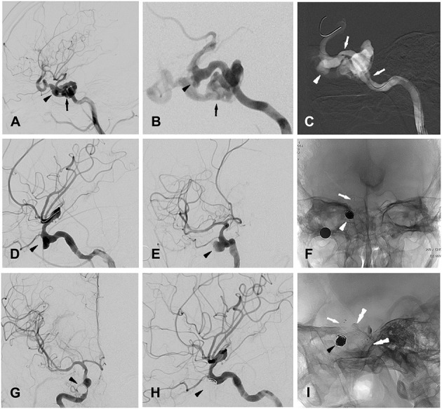
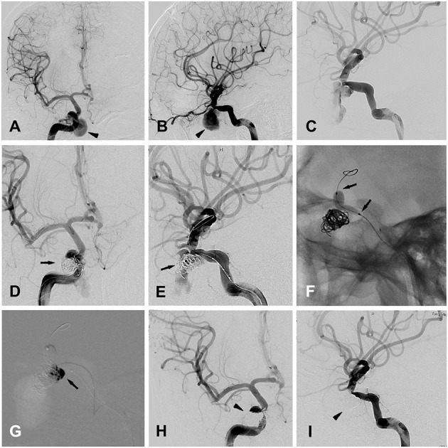
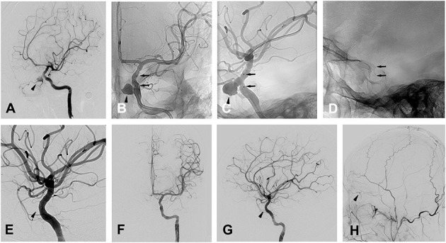
Background and Purpose: As a rare lesion secondary to brain trauma, traumatic intracranial aneurysms (TICAs) lead to high mortality and morbidity, and multiple treatment modalities have been applied for TICAs. All patients diagnosed with TICAs in our institution from 2010 to 2020 were included in the report, and their clinical features, treatment, and outcomes are described in detail. The purpose of this study is to illustrate the characteristic of different therapeutic methods of TICAs, and focus on the endovascular treatment. Methods: A total of 20 patients were included in this study. The 3 patients who declined treatment all died. Five of the other 17 patients were treated surgically, including clipping, wrapping, and trapping with or without EC-IC high-flow bypass, with only 1 case of parent artery preservation. Twelve patients underwent endovascular treatment, including bare coil embolization (1 case), stent-assisted coiling (2 cases), balloon-assisted coils/Onyx glue embolization (1 case) and covered stents (8 cases), with only 1 case of parent artery sacrifice. Results: 20 patients were included in the present study with 17 males, and the mean of age on 27 years (IQR: 22, 44 years). Eight patients presented with epistaxis, followed by 5 patients with coma, 3 patients with visual defects and 2 patients with CSF leakage. There were 18 TICAs located at the internal carotid artery (ICA); The other 2 TICAs located at pericallosal artery and A1 segment anterior cerebral artery (ACA). One case of diplopia occurred due to sacrifice of the ICA. Occlusion of the ophthalmic artery occurred in 3 patients after placement of a covered stent, with 1 patient suffering an irreversible vision decrease. None of the other patients who underwent the treatment have experienced an aggravation of their symptoms since the treatment; During the imaging follow-up, 1 case of recurrence and 1 case of endoleak occurred in this case series. Conclusions: TICAs are associated with significant morbidity and mortality, and endovascular treatment has emerged as a valuable option, which may be promising to improve the clinical outcomes due to their advantages of preserving the parent artery if occlusion of the side branch artery can be avoided.

Keywords: endovascular treatment; parent artery; side branch artery; surgical treatment; traumatic intracranial pseudoaneurysms.


Conflict of interest statement

The authors declare that the research was conducted in the absence of any commercial or financial relationships that could be construed as a potential conflict of interest.

Figures

Figure 1
Figure 1
Patient 5 (A,B) (magnified), lateral (A) and oblique (B) views of right ICA angiogram showing the traumatic carotid-cavernous fistula (CCF, indicated by black arrow) as well as one pseudoaneurysm (indicated by black arrow head) located at cavernous segment of ICA. (C) Intra-procedural road map during stent graft (white arrows) positioning for occluding the CCF, with white arrow head indicating the pseudoaneurysm. (D,E) Oblique views of right ICA angiogram showing the complete occlusion of the CCF and the remained pseudoaneurysm (indicated by black arrow head). (F) Post-treatment radioscopic anteroposterior view of the placed coils (white arrow head) and stent (white arrow) for the stent-assisted coiling procedures. (G,H) Anteroposterior (G) and lateral (H) views of a right ICA angiogram showing the completely occluded traumatic pseudoaneurysm (black arrow head). (I) post-treatment radioscopic lateral view of the placed coils (black arrow head) and the stent (white arrow) for stent-assisted coiling, as well as the covered- stent (black double-arrow heads) for treating CCF.
Figure 2
Figure 2
Patient 12 (A,B) anteroposterior (A) and lateral (B) views of the right ICA angiogram for initial diagnosis, showing a traumatic cavernous pseudoaneurysm (black arrow head). (C) The second time of right ICA angiogram before the embolization, showing the formation of the thrombus in the false cavity of the pseudoaneurysm. (D,E) Anteroposterior (A) and oblique (B) views of the right ICA angiogram, showing the placement of endovascular coils in the pseudoaneurysm (black arrows), with the embolization microcatheter directing toward the pseudoaneurysm's cavity. (F) Balloon inflation (black arrows) was accomplished with the saline/contrast mixture. (G) Intraprocedural road map during the injection of the onyx (black arrow) into the pseudoaneurysm's cavity, after the complete cover of the pseudoaneurysm's ostium by the inflated balloon. (H,I) Anteroposterior (A) and oblique (B) views of the right ICA angiogram, showing the traumatic pseudoaneurysm completely occluded by the coils and onyx embolization (black arrow heads).
Figure 3
Figure 3
Patient 13 (A) oblique views of a left ICA angiogram showing the traumatic OphA aneurysm. (B,C) radioscopic anteroposterior (B) and oblique (C) views of the implanted stent during the left ICA angiogram. Black arrow heads indicate the pseudoaneurysm close to the ophthalamic artery in (A–C), black arrows indicate the stent in (B–G), oblique (E), anteroposterior (F) and lateral (G) views of a left ICA angiogram showing the complete occlusion of the pseudoaneurysm, with black arrow head indicating the absence of the pseudoaneurysm as well as ophthalamic artery. (H) Lateral (G) view of a left ECA angiogram showing the presence of the collateral flow from ECA to OphA (black arrow head).

References

    1. Holmes B, Harbaugh RE. Traumatic intracranial aneurysms: a contemporary review. J Trauma. (1993) 35:855–60. 10.1097/00005373-199312000-00009 - DOI - PubMed
    1. Dubey A, Sung WS, Chen YY, Amato D, Mujic A, Waites P, et al. . Traumatic intracranial aneurysm: a brief review. J Clin Neurosci. (2008) 15:609–12. 10.1016/j.jocn.2007.11.006 - DOI - PubMed
    1. Cohen JE, Gomori JM, Segal R, Spivak A, Margolin E, Sviri G, et al. . Results of endovascular treatment of traumatic intracranial aneurysms. Neurosurgery. (2008) 63:476–85; discussion 85–6. 10.1227/01.NEU.0000337169.55304.A0 - DOI - PubMed
    1. Bell RS, Vo AH, Roberts R, Wanebo J, Armonda RA. Wartime traumatic aneurysms: acute presentation, diagnosis, and multimodal treatment of 64 craniocervical arterial injuries. Neurosurgery. (2010) 66:66–79; discussion 10.1227/01.NEU.0000361285.50218.A8 - DOI - PubMed
    1. Niu Y, Zhou S, Tang J, Miao H, Zhu G, Chen Z. Treatment of traumatic intracranial aneurysm: experiences at a single center. Clin Neurol Neurosurg. (2020) 189:105619. 10.1016/j.clineuro.2019.105619 - DOI - PubMed