. 2021 Jun 23:12:689269.
doi: 10.3389/fimmu.2021.689269. eCollection 2021.

HVEM Promotes the Osteogenesis of allo-MSCs by Inhibiting the Secretion of IL-17 and IFN-γ in Vγ4T Cells

Affiliations

HVEM Promotes the Osteogenesis of allo-MSCs by Inhibiting the Secretion of IL-17 and IFN-γ in Vγ4T Cells

Lei He et al. Front Immunol. .

Abstract

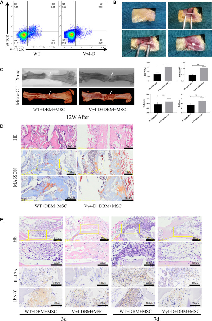
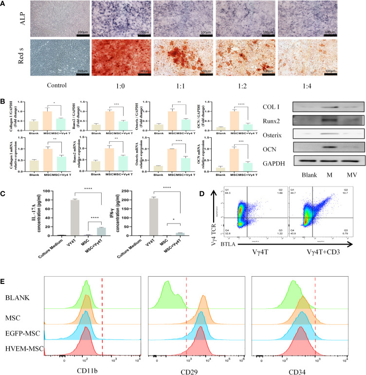
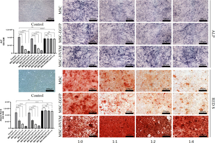
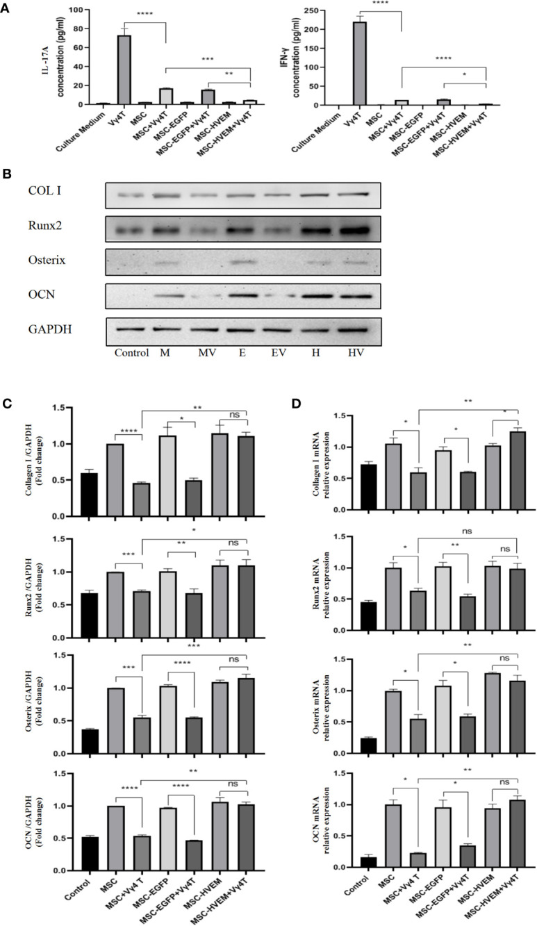
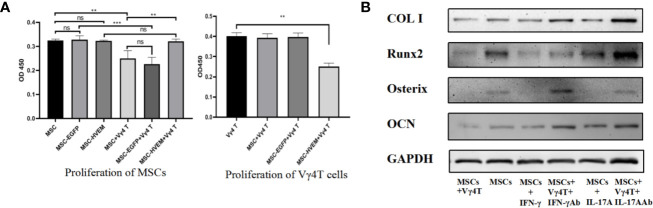
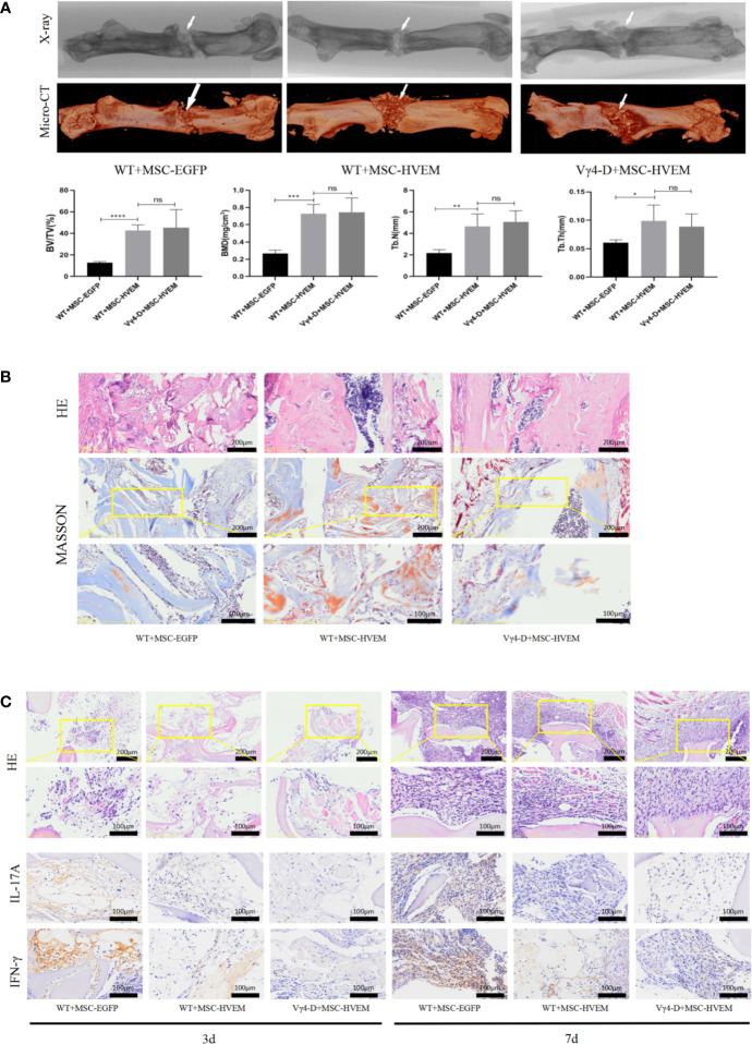
Bone defects are a common orthopaedic concern, and an increasing number of tissue-engineered bones (TEBs) are used to repair bone defects. Allogeneic mesenchymal stem cells (allo-MSCs) are used as seed cells in many approaches to develop TEB constructs, but the immune response caused by allogeneic transplantation may lead to transplant failure. V gamma 4 T (Vγ4T) cells play an important role in mediating the immune response in the early stage after transplantation; therefore, we wanted to verify whether suppressing Vγ4T cells by herpesvirus entry mediator (HVEM)/B and T lymphocyte attenuator (BTLA) signalling can promote MSCs osteogenesis in the transplanted area. In vitro experiments showed that the osteogenic differentiation of MSCs and Vγ4T cells was weakened after co-culture, and an increase in interleukin-17 (IL-17) and interferon-γ (IFN-γ) levels was detected in the culture supernatant. HVEM-transfected MSCs (MSCs-HVEM) still exhibited osteogenic differentiation activity after co-culture with Vγ4T cells, and the levels of IL-17 and IFN-γ in the co-culture supernatant were significantly reduced. In vivo experiments revealed that inflammation in the transplanted area was reduced and osteogenic repair was enhanced after Vγ4T cells were removed. MSCs-HVEM can also consistently contribute to reduced inflammation in the transplanted area and enhanced bone repair in wild-type (WT) mice. Therefore, our experiments verified that HVEM can promote the osteogenesis of allo-MSCs by inhibiting IL-17 and IFN-γ secretion from Vγ4T cells.

Keywords: HVEM-BTLA; IL-17; MSc; Tissue engineered bone; Vγ4T cells; immunomodulatory.


Conflict of interest statement

The authors declare that the research was conducted in the absence of any commercial or financial relationships that could be construed as a potential conflict of interest.

Figures

Figure 1
Figure 1
Establishment of femoral nonunion fracture model and Osteogenesis of TEB in vivo of femal Balb/c mice aged 8 weeks old. (A) Constructed Vγ4T cells free Balb/c mice (Vγ4-D). (B) Process for careting the nonunion fracture models in mice. (C) Morphological analysis of bone formation in 2 groups mice femur defect at 12 weeks after operation and Microarchitecture analyses of bone volume fraction (BV/TV) ,bone mineral density (BMD) , trabecular number of bone formation area (Tb.N) and  trabecular thickness (Tb.Th) by X-ray and 3D Reconstruction . Error bars represent mean ± SD. One-way ANOVA test was used to calculated P value (***P < 0.01, ns P > 0.05). (D) Histological analysis of newly formed bone by H&E and Masson’s Trichrome staining. (E) Infiltration of inflammatory cells and expressions of IL-17A and IFN-γ detected by immunohistochemistry at 3 days and 7 days post implantation.
Figure 2
Figure 2
Vγ4T cells inhibite the osteogenic differentiation of MSCs. (A) ALP staining and alizarin red staining show various concentrations of Vγ4T cells inhibited the osteogenic differentiation of MSCs. (B) Expression of markers of osteogenesis by MSCs with or not with Vγ4T cells(the ratio is 1:4) were measured by RT-PCR and Western blotting analyses, relative protein expression of COL I, Runx2, Osterix, and OCN by Western blotting, mRNA expression levels were tested by RT-PCR. (C) The concentration of IL-17A and IFN-γ was measured in the supernatant of co cultured cells for 3 days. (D) BTLA expression in Vγ4T cells. (E) Changes of surface markers of MSCs or HVEM- expressing MSCs (MSCs-HVEM). Error bars represent mean ± SD. One-way ANOVA test was used to calculated P value (*P < 0.05, **P < 0.01, ***P < 0.001, ****P < 0.0001).
Figure 3
Figure 3
Vγ4T cells co-culture with MSCs or MSCs-HVEM then induced in osteogenic medium and ALP staining and alizarin red staining show various concentrations of Vγ4T cells inhibited the osteogenic differentiation of MSCs but MSCs-HVEM could reverse the inhibition of Vγ4T cells. Error bars represent mean ± SD. One-way ANOVA test was used to calculated P value (****P < 0.0001, ns P > 0.05).
Figure 4
Figure 4
Vγ4T cells co-culture with MSCs or MSCs-HVEM then induced in osteogenic medium. (A) MSC can inhibit IL-17A and IFN-γ secretion from purified Vγ4T cells cells, the inhibition ability of MSCs-HVEM was stronger than that of MSC cells. (B, C) The Expression of markers of osteogenesis such as COL I, Runx2, Osterix, and OCN by MSCs or MSCs-HVEM  with or not with Vγ4T cells(the ratio is 1:4) were measured by Western blotting analyses, the osteogenic differentiation ability of MSC decreased when co-culture with Vγ4T cells but MSCs-HVEM can counter or even reverse that trend .MSCs (M),MSCs + Vγ4T cells (MV), MSCs-EGFP (E), MSCs-EGFP + Vγ4T cells (EV), MSCs-HVEM (H), MSCs-HVEM + Vγ4T cells (HV). (D) mRNA expression levelsof COL I, Runx2, Osterix, and OCN were tested by RT-PCR after 9 days of MSCs or MSCs-HVEM with or not with Vγ4T cells (the ratio is 1:4, relative to expression in MSCs showed the same trend. Error bars represent mean ± SD. One-way ANOVA test was used to calculated P value (*P < 0.05, **P < 0.01, ***P < 0.001, ****P < 0.0001, ns P > 0.05).
Figure 5
Figure 5
The effect of Vγ4T cells on MSCs proliferation and osteogenic differentiation. (A) The proliferation of cell was measured by using CCK-8 assay on 3rd day. Absorbance values at 450 nm for each group are shown when MSCs or MSCs-HVEM co-culture with or not with Vγ4T cells. Data are reported as means ± SD. * 0.01 ≤ P ≤ 0.05, ** P ≤ 0.01, *** P ≤ 0.001 (One Way ANOVA). (B) The Expression of markers of osteogenesis such as COL I, Runx2, Osterix, and OCN by MSCs with Vγ4T cells (the ratio is 1:4) treated with IFN-γ, IFN-γ antibody (IFN-γ Ab) , IL-17A, IL-17A antibody (IL-17A Ab) are shown. Error bars represent mean ± SD. One-way ANOVA test was used to calculated P value (**P < 0.01, ***P < 0.001, ns P > 0.05).
Figure 6
Figure 6
Osteogenic ability and Local immune response of TEB constructed by MSCHVEM in vivo. (A) Both WT+MSCHVEM groups and VY4-D+MSCHVEM groups show higher quality of bone formation than that of WT+MSC in BV/TV, BMD, Tb.N, Tb.Th, but the quality of bone formation of between WT+MSCHVEM groups and VY4-D+ MSCHVEM groups had no statistics difference (P > 0.05). (B) Histological analysis of newly formed bone by H&E and Masson’s Trichrome staining in 3 groups. (C) Infiltration of inflammatory cells and expressions of IL-17A and IFN-γ detected by immunohistochemistry at 3 days and 7 days post implantation. Error bars represent mean ± SD. One-way ANOVA test was used to calculated P value (*P < 0.05, **P < 0.01, ***P < 0.001, ****P < 0.0001, ns P > 0.05).

Similar articles

Cited by

References

    1. Dai F, Shi D, He W, Wu J, Luo G, Yi S, et al. . hCTLA4-Gene Modified Human Bone Marrow-Derived Mesenchymal Stem Cells as Allogeneic Seed Cells in Bone Tissue Engineering. Tissue Eng (2006) 12(9):2583–90. 10.1089/ten.2006.12.2583 - DOI - PubMed
    1. Tang D, Tare R, Yang L, Williams D, Ou K, Oreffo R. Biofabrication of Bone Tissue: Approaches, Challenges and Translation for Bone Regeneration. Biomaterials (2016) 83:363–82. 10.1016/j.biomaterials.2016.01.024 - DOI - PubMed
    1. Niemeyer P, Fechner K, Milz S, Richter W, Suedkamp N, Mehlhorn A, et al. . Comparison of Mesenchymal Stem Cells From Bone Marrow and Adipose Tissue for Bone Regeneration in a Critical Size Defect of the Sheep Tibia and the Influence of Platelet-Rich Plasma. Biomaterials (2010) 31(13):3572–9. 10.1016/j.biomaterials.2010.01.085 - DOI - PubMed
    1. Xi J, Yan X, Zhou J, Yue W, Pei X. Mesenchymal Stem Cells in Tissue Repairing and Regeneration: Progress and Future. Burns Trauma (2013) 1(1):13–20. 10.4103/2321-3868.113330 - DOI - PMC - PubMed
    1. Hu P, Yang Q, Wang Q, Shi C, Wang D, Armato U, et al. . Mesenchymal Stromal Cells-Exosomes: A Promising Cell-Free Therapeutic Tool for Wound Healing and Cutaneous Regeneration. Burns Trauma (2019) 7:38. 10.1186/s41038-019-0178-8 - DOI - PMC - PubMed

Publication types