Review
. 2021 Jun 23:12:705920.
doi: 10.3389/fimmu.2021.705920. eCollection 2021.

Metabolic Control of Smoldering Neuroinflammation

Affiliations
Review

Metabolic Control of Smoldering Neuroinflammation

Luca Peruzzotti-Jametti et al. Front Immunol. .

Abstract

Compelling evidence exists that patients with chronic neurological conditions, which includes progressive multiple sclerosis, display pathological changes in neural metabolism and mitochondrial function. However, it is unknown if a similar degree of metabolic dysfunction occurs also in non-neural cells in the central nervous system. Specifically, it remains to be clarified (i) the full extent of metabolic changes in tissue-resident microglia and infiltrating macrophages after prolonged neuroinflammation (e.g., at the level of chronic active lesions), and (ii) whether these alterations underlie a unique pathogenic phenotype that is amenable for therapeutic targeting. Herein, we discuss how cell metabolism and mitochondrial function govern the function of chronic active microglia and macrophages brain infiltrates and identify new metabolic targets for therapeutic approaches aimed at reducing smoldering neuroinflammation.

Keywords: immunometabolism; macrophages; metabolism; microglia; mitochondria; progressive multiple sclerosis; smoldering inflammation.


Conflict of interest statement

SP is co-founder, CSO, and shareholder (>5%) of CITC Ltd. and iSTEM Therapeutics and co-founder and Non-Executive Director at Asitia Therapeutics; LP-J is a shareholder of CITC Ltd. The remaining authors declare that the research was conducted in the absence of any commercial or financial relationships that could be construed as a potential conflict of interest.

Figures
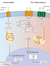
Figure 1
Figure 1
Glucose metabolism in microglia under homeostatic and inflammatory conditions. Under homeostatic conditions, extracellular glucose is transported into microglial cells through specialized glucose transporters, where it is converted into pyruvate through cytoplasmic glycolysis. Pyruvate is then actively transported across the mitochondrial membrane to drive the TCA cycle. The energy and metabolites produced in the TCA cycle can then support the expression of the homeostatic microglial genes P2ry12 and Tmem119, which facilitate microglial functions of synaptic pruning and immune surveillance. In pro-inflammatory conditions, microglia have a broken TCA cycle and increase the expression of membrane transporters to facilitate the uptake of glucose and glutamine, thus driving enhanced glycolysis and glutaminolysis. Glycolysis is supported by increased expression of the rate-limiting enzymes of glycolysis HK2 and PFKBP3. This leads to the increased generation of lactate and ATP to compensate for the broken TCA cycle, and shunting of metabolites into the pentose phosphate pathway. The increased glycolysis is sustained by the activation of nuclear transcription factors HIF1α and mTOR that support the synthesis and production of cytokines for secretion. Green arrows = homeostatic effects. Red arrows = pro-inflammatory effects. GLUT, glucose transporter; TCA, tricarboxylic acid cycle; GLT, glutamate transporter; HK2, hexokinase 2; PFKBP3, 6-phosphofructo-2-kinase/fructose-2,6-biphosphate-3; HIF1α, hypoxia inducible factor 1 α; ATP, adenosine triphosphate; mTOR, mechanistic target of rapamycin; TNFα, tumor necrosis factor alpha; Il1β, interleukin-1β; Il6, interleukin-6.
Figure 2
Figure 2
Lipid metabolism in microglia under homeostatic and inflammatory conditions. In microglia, CD36 and TREM2 play a key role in the response to extracellular lipids. CD36 promotes lipid-responsive signalling pathways (like the PPARγ and LXR pathways), which in turn increase FAO and further upregulate Cd36. TREM2 activation (via ligands such as ApoE) results in the suppression of homeostatic microglial genes (P2ry12, Tmem119, and Cx3cr1), the activation of mTOR signalling, and the upregulation of lipid processing genes (such as Apoe, Lpl, and Fabp5). Despite mTOR increasing both FAS (through the cleavage and activation of SREBP-1) and glycolysis (which are canonically associated with a pro-inflammatory activation of myeloid cells), it appears that the role of TREM2 is to support correct lipid metabolism. In fact,TREM2 deficient microglia show the formation of intracellular cholesterol crystals that activate the inflammasome pathway. On the contrary, the downstream gene Fabp5 seems to play a key role in determining the pro-inflammatory activation of myeloid cells, possibly via inhibition of PPARγ signalling and FAO. In pro-inflammatory microglia, a broken TCA cycle is coupled with an upregulated mitochondrial CIC, which increases citrate export from the mitochondria to the cytosol, where it is converted into acetyl-CoA for FAS by ACLY. The resultant increase in FAS supports the expansion of the ER and Golgi, and the increased production of pro-inflammatory cytokines. Green arrows = homeostatic effects. Red arrows = pro-inflammatory effects. PPARγ, peroxisome proliferator-activated receptor γ; LXR, liver X receptor; FAO, fatty acid oxidation; mTOR, mechanistic target of rapamycin; FAS, fatty acid synthesis; ACLY, ATP citrate lyase; CIC, citrate carrier; TCA, tricarboxylic acid cycle; SREBP-1, Sterol regulatory element binding protein 1; TYROBP, TYRO protein tyrosine kinase-binding protein; TREM2, Triggering receptor expressed on myeloid cells 2; Lpl, Lipoprotein Lipase; P2ry12, Purinergic Receptor P2Y12; Tmem119, Transmembrane Protein 119, Cx3Cr, C-X3-C Motif Chemokine Receptor 1.
Figure 3
Figure 3
Pro-inflammatory conditions lead to morphological and functional mitochondrial alterations of microglia. Microglia in homeostatic conditions have intact mitochondria with a functioning TCA cycle and conserved fusion of the mitochondrial network through OPA1 and MFN1/2 activities. Succinate signalling through SUCNR1 in homeostasis is presumably low, but its signalling functions in resting microglia are currently undefined. In pro-inflammatory conditions, mitochondria undergo DRP1-mediated fission and fragmentation, and show two breaks in the TCA cycle that lead to the intracellular accumulation of specific metabolites (such as succinate, citrate, and itaconate). Succinate accumulation within mitochondria can drive RET, which produces excessive mtROS through complex I. This mitochondrial dysfunction creates a pseudohypoxic state that leads to the stabilization of HIF1α and enhances cytokine production and secretion. Both fragmented mitochondria and succinate can be released into the extracellular environment where succinate can signal in an autocrine or paracrine manner via SUCNR1, thus modulating both anti-inflammatory and pro-inflammatory effects. Green arrows = anti-inflammatory effects; red arrows = pro-inflammatory effects. SUCNR1, succinate receptor 1; TCA, tricarboxylic acid cycle; OPA1, optic atrophy 1; MFN1/2, mitofusin 1/2; DRP1, dynamin-related protein 1; CI, complex I; CII, complex II; CIII, complex III; mtROS, mitochondrial reactive oxygen species; RET, reverse electron transport; ATP, adenosine triphosphate; HIF1α, hypoxia inducible factor 1 α; mtROS, mitochondrial reactive oxygen species; TNFα, tumor necrosis factor alpha; Il1β, interleukin-1β; Il6, interleukin-6.

References

    1. Zhu J, Thompson CB. Metabolic Regulation of Cell Growth and Proliferation. Nat Rev Mol Cell Biol (2019) 20:436–50. 10.1038/s41580-019-0123-5 - DOI - PMC - PubMed
    1. Chipuk JE, Mohammed JN, Gelles JD, Chen Y. Mechanistic Connections Between Mitochondrial Biology and Regulated Cell Death. Dev Cell (2021) 56(9):1221–33. 10.1016/j.devcel.2021.03.033 - DOI - PMC - PubMed
    1. Balaban RS. Regulation of Oxidative Phosphorylation in the Mammalian Cell. Am J Physiol (1990) 258:C377–89. 10.1152/ajpcell.1990.258.3.C377 - DOI - PubMed
    1. Spinelli JB, Haigis MC. The Multifaceted Contributions of Mitochondria to Cellular Metabolism. Nat Cell Biol (2018) 20:745–54. 10.1038/s41556-018-0124-1 - DOI - PMC - PubMed
    1. Miyazawa H, Aulehla A. Revisiting the Role of Metabolism During Development. Dev (Cambridge England) (2018) 145(19):dev131110. 10.1242/dev.131110 - DOI - PubMed

Publication types

LinkOut - more resources