Review
. 2022 Apr;12(4):1591-1623.
doi: 10.1016/j.apsb.2021.06.016. Epub 2021 Jul 2.

Recent advances in developing small-molecule inhibitors against SARS-CoV-2

Affiliations
Review

Recent advances in developing small-molecule inhibitors against SARS-CoV-2

Rong Xiang et al. Acta Pharm Sin B. 2022 Apr.

Abstract

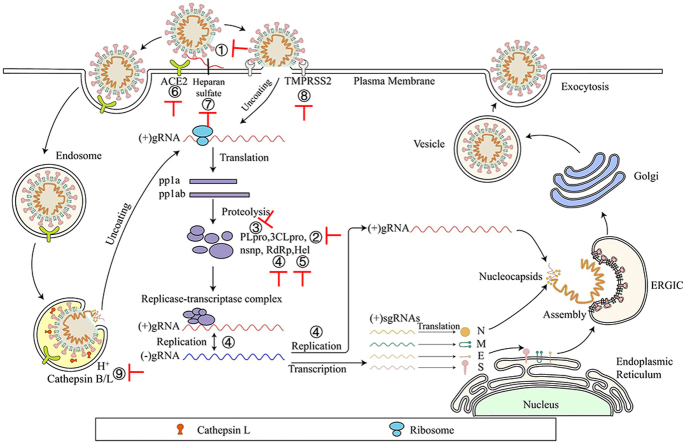
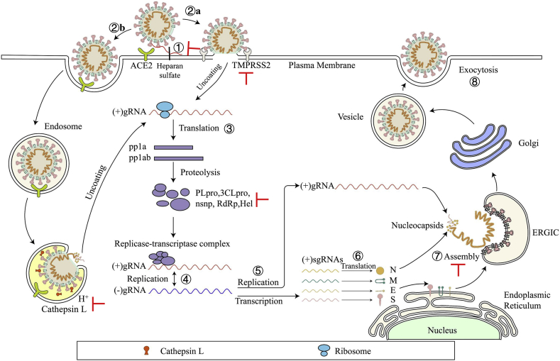
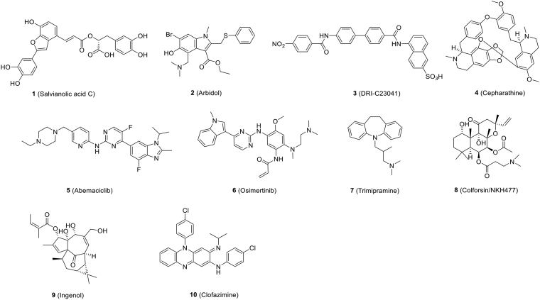
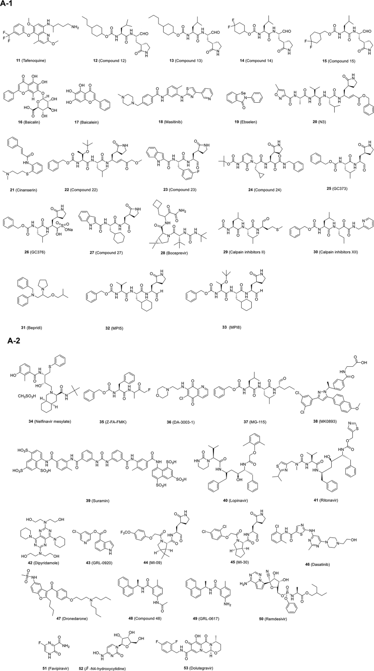
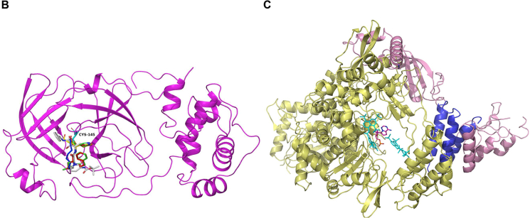
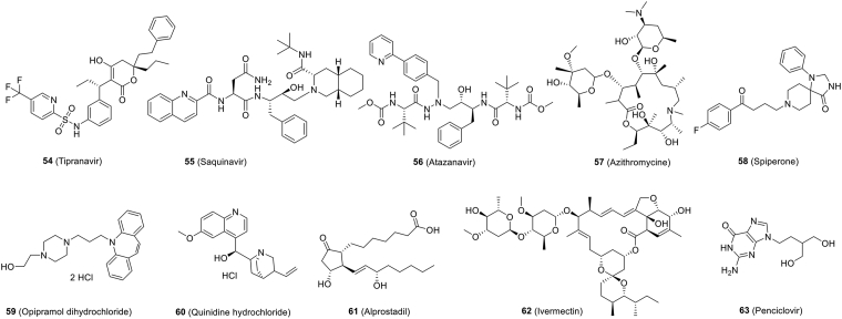
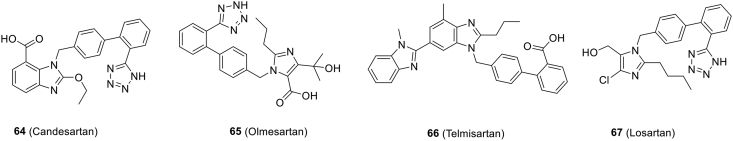
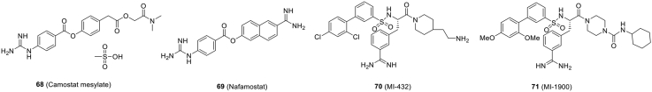
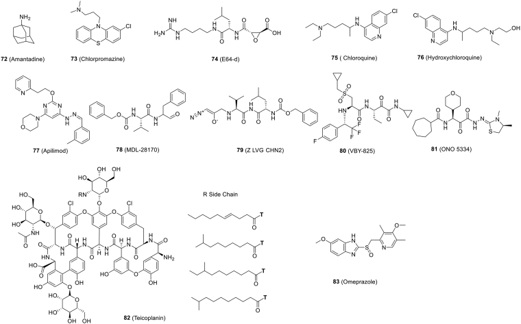
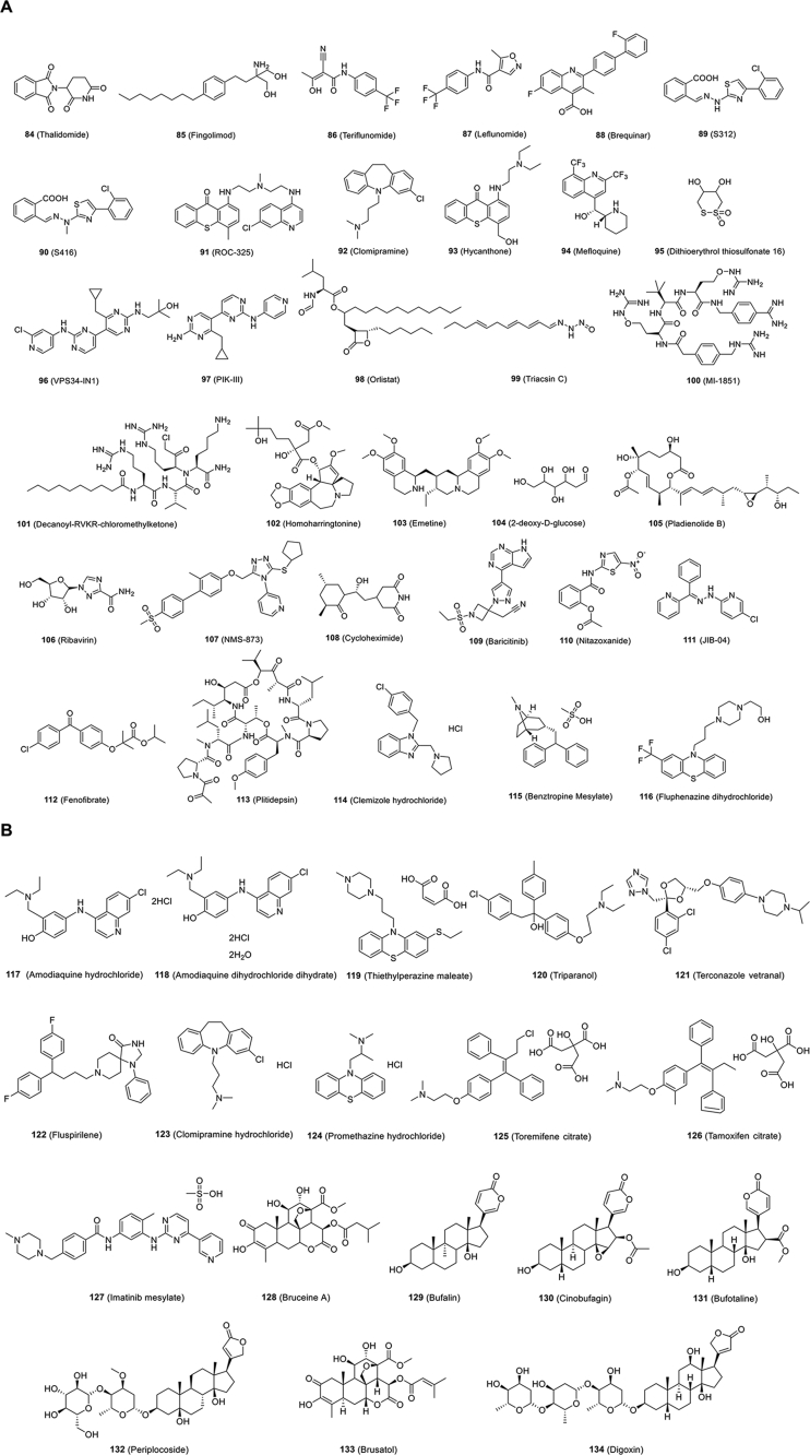
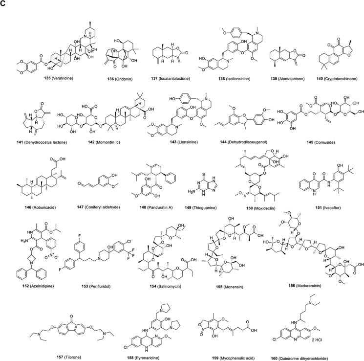
The COVID-19 pandemic caused by the novel SARS-CoV-2 virus has caused havoc across the entire world. Even though several COVID-19 vaccines are currently in distribution worldwide, with others in the pipeline, treatment modalities lag behind. Accordingly, researchers have been working hard to understand the nature of the virus, its mutant strains, and the pathogenesis of the disease in order to uncover possible drug targets and effective therapeutic agents. As the research continues, we now know the genome structure, epidemiological and clinical features, and pathogenic mechanism of SARS-CoV-2. Here, we summarized the potential therapeutic targets involved in the life cycle of the virus. On the basis of these targets, small-molecule prophylactic and therapeutic agents have been or are being developed for prevention and treatment of SARS-CoV-2 infection.

Keywords: COVID-19; Prophylactic; SARS-CoV-2; Small-molecule inhibitors; Therapeutic.


Figures

Image 1
Graphical abstract
Figure 1
Figure 1
Schematic diagram of genome composition and particle structure of SARS-CoV-2. SARS-CoV-2 genome consists of open reading frames (ORFs) expressing structural proteins including spike protein (S), envelope protein (E), membrane protein (M), and nucleocapsid protein (N), and ORFs1a, 1 b, etc. Expressing non-structural and accessory proteins. Among them, N protein and (+) ssRNA form nucleocapsids with three other structural proteins to form mature viral particles.
Figure 2
Figure 2
Life cycle of SARS-CoV-2. SARS-CoV-2 first binds, via its S protein, to the receptor ACE2 on the target cell (①). Then, the virus must gain access to the host cell cytosol through plasma (②a) or endosomal membrane fusion (②b). This is assisted by activation of S protein by TMPRRS2 (②a) or cathepsin B/L (②b), followed by fusion of the viral and cellular membranes. The viral genome is released, uncoated and translated into viral replicase polyproteins pp1a and 1 ab (③), which are then cleaved into nonstructural proteins (nsps) by viral proteinases as papain-like protease (PLpro) and 3C-like protease (3CLpro). Many of these nsps as RNA-dependent RNA polymerase (RdRp) or Helicase (Hel) then assemble into the replicase–transcriptase complex which replicates the (+)-sense genomic RNA ((+) gRNA). (−)-sense genomic RNA ((−) gRNA) is synthesized and used as a template to form (+) gRNA) and subgenomic RNAs (sgRNAs) (⑤). The viral structural proteins, S, E, and M are translated from sgRNAs (⑥) and inserted into the endoplasmic reticulum (ER), from where they are transported to the ER–Golgi intermediate compartment (ERGIC) to interact with the (+) gRNA-encapsidated N proteins and assemble into viral particles (⑦). The budded vesicles containing mature viral particles are then transported to the cell surface for release after maturation in the Golgi bodies (⑧). Possible targets for inhibitors are marked in red.
Figure 3
Figure 3
Structural regions and fusion mechanism of SARS-CoV-2 S protein. (A) The functional regions in SARS-CoV-2 S protein include SP (signal peptide, light yellow), RBD (receptor-binding domain; light green), FP (fusion peptide; light blue), HR1 (heptad repeat 1; gray-blue), HR2 (heptad repeat 2; flesh), TM (transmembrane; grass green), and CP (cytoplasmic; purple). (B) SARS-CoV-2 S protein fusion pathway base on class I fusion protein. The S protein starts in the native state and undergoes priming of the S1 subunit by relevant proteases to achieve the prefusion state. Subsequent triggering by relevant proteases will enable the FP to insert in the host membrane and allow the S protein to form the prehairpin intermediate. The prehairpin begins to fold back on itself due to HR1 and HR2 interactions forming the 6-HB, and eventual postfusion stable states. During the S protein foldback, the two membranes will approach each other until the outer leaflets merge (hemifusion) and eventually the inner leaflets merge.
Figure 4
Figure 4
Chemical structures of small-molecule inhibitors that inhibit SARS-CoV-2 entry.
Figure 5
Figure 5
SARS-CoV-2 replication inhibitors. (A) Chemical structures of SARS-CoV-2 replication inhibitors. (B) Overall views of the 3CLpro-N3 (yellow) complex overlapped with carmofur (blue), 13 b (silver), GC373 (red) and GC376 (green) (PDB ID: 7BQY, 7BUY, 6Y2F, 6WTJ and 6WTK), and amino residue Cys145 was shown as cyan. (C) Overall views of the RdRp-suramin (cyan) complex overlapped with the remdesivir (magenta)-bound RdRp structure (PDB ID: 7D4F and 7BV2). nsp 12 was shown as yellow and accessary subunits nsp 7 and nsp 8 were shown as blue and pink.
Figure 5
Figure 5
SARS-CoV-2 replication inhibitors. (A) Chemical structures of SARS-CoV-2 replication inhibitors. (B) Overall views of the 3CLpro-N3 (yellow) complex overlapped with carmofur (blue), 13 b (silver), GC373 (red) and GC376 (green) (PDB ID: 7BQY, 7BUY, 6Y2F, 6WTJ and 6WTK), and amino residue Cys145 was shown as cyan. (C) Overall views of the RdRp-suramin (cyan) complex overlapped with the remdesivir (magenta)-bound RdRp structure (PDB ID: 7D4F and 7BV2). nsp 12 was shown as yellow and accessary subunits nsp 7 and nsp 8 were shown as blue and pink.
Figure 6
Figure 6
Chemical structures of other small-molecule inhibitors against SARS-CoV-2.
Figure 7
Figure 7
Chemical structures of small-molecule inhibitors targeting ACE2.
Figure 8
Figure 8
Chemical structures of small-molecule inhibitors targeting TMPRSS2.
Figure 9
Figure 9
Chemical structures of small-molecule inhibitors targeting cathepsin B/L.
Figure 10
Figure 10
Chemical structures of other small-molecule inhibitors targeting host.
Figure 10
Figure 10
Chemical structures of other small-molecule inhibitors targeting host.

References

    1. Zhu N., Zhang D., Wang W., Li X., Yang B., Song J., et al. A novel coronavirus from patients with pneumonia in China, 2019. N Engl J Med. 2020;382:727–733. - PMC - PubMed
    1. Wu F., Zhao S., Yu B., Chen Y.M., Wang W., Song Z.G., et al. Author correction: a new coronavirus associated with human respiratory disease in China. Nature. 2020;580 e7. - PMC - PubMed
    1. Jiang S., Shi Z., Shu Y., Song J., Gao G.F., Tan W., et al. A distinct name is needed for the new coronavirus. Lancet. 2020;395:949. - PMC - PubMed
    1. Kim J.M., Chung Y.S., Jo H.J., Lee N.J., Kim M.S., Woo S.H., et al. Identification of coronavirus isolated from a patient in Korea with COVID-19. Osong Public Health Res Perspect. 2020;11:3–7. - PMC - PubMed
    1. Zhou P., Yang X.L., Wang X.G., Hu B., Zhang L., Zhang W., et al. A pneumonia outbreak associated with a new coronavirus of probable bat origin. Nature. 2020;579:270–273. - PMC - PubMed