

Giant Cell Tumor (GCT) of the Third Metatarsal in an Elderly Patient: A Rare Case Report
- PMID: 34249798
- PMCID: PMC8241254
- DOI: 10.13107/jocr.2021.v11.i03.2074
Giant Cell Tumor (GCT) of the Third Metatarsal in an Elderly Patient: A Rare Case Report
Abstract
Introduction: Giant cell tumour (GCT) is a benign osteolytic, locally aggressive lesion. Seen in young adults at the epiphysis. The most common site is long bones (85-90%). GCT of the metatarsal in elderly patients is very rare.
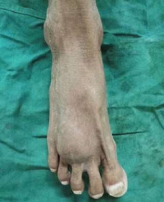
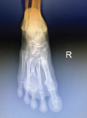
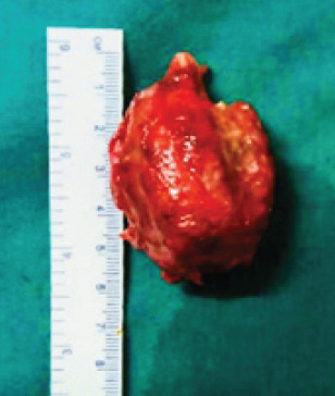
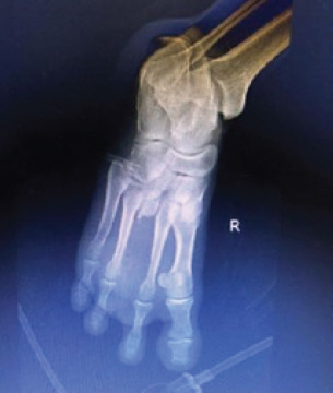
Case report: A 60-year-old male came with complaints of pain and swelling over right foot dorsal aspect since for the last past one 1 year. There was no history of trauma. X-ray foot showed an osteolytic lesion in the right third metatarsal with thinning of the cortex. MRI and fine-needle aspiration cytology confirmed the diagnosis of GCT. The patient was managed by excision with the 3rd ray amputation. At present, 1.5 years follow-up, the patient is having no pain, difficulty in walking and no evidence of clinical and radiological recurrence .
Conclusion: Giant cell tumours could also present at uncommon sites, and they should be considered in the differential diagnosis of lytic lesions of the metatarsals. Excision with ray amputation of the involved metatarsal helps in complete removal of the lesion and helps in early weight-bearing. This is the viable alternative treatment option in managing the metatarsal GCT in elderly patients.
Keywords: Giant cell tumour; ray amputation; third metatarsal.
Copyright: © Indian Orthopaedic Research Group.
Conflict of interest statement
Conflict of Interest: Nil
Figures


References
-
- Unni KK. Dahlin's Bone Tumors:General Aspects and Data on 11,087 Cases. 5th ed. Philadelphia, PA: Lippincott-Raven; 1996. pp. 263–83.
-
- Mohan V, Gupta SK, Sharma OP, Varma DN. Giant cell tumor of short tubular bones of the hands and feet. Indian J Radiol. 1980;34:1–17.
-
- Mendicino SS. Giant cell tumor of the first metatarsal bone en bloc resection with autogenous middle fibular strut graft. J Foot Ankle Surg. 1993;32:405–10. - PubMed
-
- Bertoni F, Bacchini P, Staals EL. Malignancy in giant cell tumor of bone. Cancer. 2003;97:2520–9. - PubMed
-
- Huvos AG. Bone Tumors:Diagnosis, Treatment and Prognosis. Philadelphia, PA: Saunders; 1991. pp. 429–67.
Publication types
LinkOut - more resources
Full Text Sources
Miscellaneous