. 2021 Jun 25:9:690358.
doi: 10.3389/fbioe.2021.690358. eCollection 2021.

Detrimental Effect of Various Preparations of the Human Amniotic Membrane Homogenate on the 2D and 3D Bladder Cancer In vitro Models

Affiliations

Detrimental Effect of Various Preparations of the Human Amniotic Membrane Homogenate on the 2D and 3D Bladder Cancer In vitro Models

Aleksandar Janev et al. Front Bioeng Biotechnol. .

Abstract

Despite being among the ten most common cancers with high recurrence rates worldwide, there have been no major breakthroughs in the standard treatment options for bladder cancer in recent years. The use of a human amniotic membrane (hAM) to treat cancer is one of the promising ideas that have emerged in recent years. This study aimed to investigate the anticancer activity of hAM homogenate on 2D and 3D cancer models. We evaluated the effects of hAM homogenates on the human muscle invasive bladder cancer urothelial (T24) cells, papillary cancer urothelial (RT4) cells and normal porcine urothelial (NPU) cells as well as on human mammary gland non-tumorigenic (MCF10a) cells and low-metastatic breast cancer (MCF7) cells. After 24 h, we observed a gradual detachment of cancerous cells from the culture surface, while the hAM homogenate did not affect the normal cells. The most pronounced effect hAM homogenate had on bladder cancer cells; however, the potency of their detachment was dependent on the treatment protocol and the preparation of hAM homogenate. We demonstrated that hAM homogenate significantly decreased the adhesion, growth, and proliferation of human bladder invasive and papillary cancer urothelial cells and did not affect normal urothelial cells even in 7-day treatment. By using light and electron microscopy we showed that hAM homogenate disrupted the architecture of 2D and 3D bladder cancer models. The information provided by our study highlights the detrimental effect of hAM homogenate on bladder cancer cells and strengthens the idea of the potential clinical application of hAM for bladder cancer treatment.

Keywords: 2D and 3D in vitro models; cancer; cell cycle; light and electron microscopy; proliferation; urothelium.


Conflict of interest statement

The authors declare that the research was conducted in the absence of any commercial or financial relationships that could be construed as a potential conflict of interest.

Figures

FIGURE 1
FIGURE 1
Human amniotic membrane (hAM) homogenate preparation protocol. (A) Separation of the hAM from human chorionic membrane (hCM). (B) Washing the hAM in sterile PBS. (C) Measuring the volume of hAM pieces. (D) Addition of an appropriate culture medium to the hAM pieces in the ratio of 1:4. (E) Filtration of hAM homogenate through sterile nylon membrane filter with pore size <1 mm, after completed homogenization. (F) Cryopreserved hAM homogenate is used for further experiments.
FIGURE 2
FIGURE 2
Human amniotic membrane (hAM) homogenate causes detachment of various cancer cell types but not of normal cells. (A,C,E,G,I) After 24-h incubation with an appropriate culture medium without hAM homogenate, T24, RT4, NPU, MCF7, and MCF10a cells remained firmly attached to the culture surface. (B,D,H) 24-h incubation with hAM homogenate resulted in significant detachment of cancer T24, RT4, and MCF7 cells, (F,J) but not of normal NPU and MCF10a cells. The most presentative images are shown. (K) The percentage of surface area covered after 24-h treatment with hAM homogenate. The data are presented as mean surface area coverage ± SEM (SEM, the standard error of the mean). Data were obtained from three independent experiments, each performed with a different biological sample of hAM. Within each experiment, three technical replicates were performed. Scale bars: 100 μm. *p < 0.05.
FIGURE 3
FIGURE 3
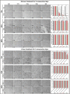
Different hAM homogenate preparations cause detachment of cancer urothelial cells in a time-dependent manner. (A1–A6) Confluent cultures of T24, RT4, and NPU cells incubated with an appropriate culture medium without hAM homogenate remained attached to the culture surface after the 72-h treatment period. (B1–E2) 72-h treatment with different hAM homogenate preparations resulted in a significant detachment of T24 cells. (B3–E4) Only hAM homogenate prepared with Russell Hobbs caused detachment of RT4 cells after the 72-h treatment period. (B5–E6) Different hAM homogenate preparations did not cause any detachment of NPU cells after the 72-h treatment period. (I1–I6) Confluent cultures of T24, RT4, and NPU cells incubated with an appropriate culture medium without hAM homogenate remained attached to the culture surface on the third day of the 2-h treatment period. (J1–M2) Different hAM homogenate preparations caused detachment of T24 cells on the third day of the 2-h treatment period. (J3–M4) Confluent cultures of RT4 cells incubated with different hAM homogenate preparations remained attached to the culture surface on the third day of the 2-h treatment period. (J5–M6) Different hAM homogenate preparations did not induce detachment of NPU cells in three consecutive days of 2-h treatment. (F,N) The percentage of surface area covered with T24 cells after 24- and 2-h treatment for three consecutive days. (G,O) The percentage of surface area covered with RT4 cells after 24- and 2-h treatment for three consecutive days. (H,P) The percentage of surface area covered with NPU cells after 24- and 2-h treatment for three consecutive days. The quantified data here are presented as mean surface area coverage ± SEM. Data were obtained from three independent experiments, each performed with a different biological sample of hAM. Within each experiment, three technical replicates were performed. Scale bars: 100 μm. *p < 0.05.
FIGURE 4
FIGURE 4
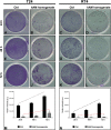
Human amniotic membrane (hAM) homogenate inhibits the cell attachment of T24 and RT4 cells and hinders their growth dynamics. (A,B) hAM homogenate significantly reduced the ability of T24 cells to attach to the culture surface after 24-h incubation. (C,D) hAM homogenate significantly reduced the ability of RT4 cells to attach to the culture surface after 24-h incubation. (E,F,I,J) hAM homogenate strongly inhibited the growth dynamics of the adhered T24 cells 48- and 72-h after the cell seeding. (G,H,K,L) hAM homogenate strongly inhibited the growth dynamics of the adhered RT4 cells 48- and 72-h after the cell seeding. (M,N) Quantitative analysis of the relative intensity of adherent T24 and RT4 cells. The regression line of the untreated cells (black dashed line) has a steeper upward tilt in comparison with the regression line of cells treated with hAM homogenate (red dashed line). The quantified data here is presented as a mean relative intensity ± standard error of the mean (SEM). Data were obtained from three distinct experiments, each performed with a different biological sample of hAM. Within each experiment, three technical replicates were carried. *p < 0.05.
FIGURE 5
FIGURE 5
Human amniotic membrane (hAM) homogenate decreases proliferation of T24 and RT4 cells and downregulates the expression of cyclin D1 in T24 cells. (A) The proliferation of T24 cells was decreased after 24, 48, and 72 h of treatment with hAM homogenate. (B) The proliferation of RT4 cells was decreased after 24, 48, and 72 h of treatment with hAM homogenate. (C,D) Western blot analysis of cyclin D1 and α-tubulin expression in T24 and RT4 cells treated with culture medium (ctrl) or hAM homogenate. The western blot analysis showed significant decrease in the expression levels of cyclin D1 after 24-h treatment with hAM homogenate in T24 cells. In RT4 cells, on the other hand, hAM homogenate induced slight but not significant decrease of cyclin D1 expression. All data shown here were obtained from at least three independent replications of experiments using three biological samples of hAM; each experiment was performed in two technical repeats for each condition. Bars represent mean ± SEM. *p < 0.05.
FIGURE 6
FIGURE 6
Structure of hAM and hAM homogenate. (A) Intact hAM is comprised of hAEC and hAM stroma, which is further divided into the compact layer, hAMSC layer and spongy layer. (B,B’,D,E) hAEC are of cuboidal form, are well connected and exhibit numerous microvilli at the apical surface. (C,C’,F,G) The fibers that form the extracellular matrix of hAM’s stroma are tightly interwoven. (D) The hAM stroma is further divided into the compact layer (CL), hAMSC layer (hAMSCL), and spongy layer (SL). (H–J) The hAM homogenate applied to T24 cells. The hAM homogenate (white asterisks) consisted of the tightly interwoven fibers of the extracellular matrix of hAM’s stroma and hAM-derived cells. The hAM homogenate applied to T24 cells adhered to the cell surface. The blue rectangles in panels (B,C) mark the areas enlarged in panels (B’,C’). The green rectangle marks the area enlarged in panel (H’); the red rectangle marks the area enlarged in panel (H”). White framed areas in panels (I,J) mark the areas enlarged in panels (I’,J’). (A) 100 μm, (B,C) 10 μm, (H) 50 μm, (H’) 25 μm, (D) 6 μm, (B’,C,E,H”,I,J) 2 μm, (J’) 600 nm, (F,G,I’) 400 nm.
FIGURE 7
FIGURE 7
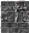
The hAM homogenate adheres to the surface of T24 and RT4 cells. (A–B’) The T24 cells incubated in culture medium for 24 or 72 h had mesenchymal morphology and there were large intercellular spaces between the cells. (C–D’) The 24- and 72-h incubation in hAM homogenate did not significantly affect the morphology of T24 cells. The hAM homogenate adhered to the surface of T24 cells and a large portion of cells were covered with it. (E–F’) The RT4 cells incubated in culture medium for 24 or 72 h had epithelial morphology and were well connected. (G–H’) The 24- and 72-h incubation in hAM homogenate did not significantly affect the morphology of RT4 cells. The hAM homogenate adhered to the surface of RT4 cells and a large portion of cells were covered with it. (I–J’) The NPU cells incubated in culture medium for 24 or 72 h retained the apical topography of well-differentiated normal urothelial cells. The apical plasma membrane appeared as ropy and rounded ridges, and rarely microridges. (K–L’) The 24- and 72-h incubation in hAM homogenate did not significantly affect the morphology of NPU cells and the hAM homogenate did not adhere to the surface of NPU cells. All data shown here were obtained from at least three independent replications of experiments using three biological samples of hAM; each experiment was performed in 1–2 technical repeats for each condition. Red arrows–hAM homogenate. Frames in panels (A–L) mark enlarged areas shown in panels (A–L’). Scale bars: (A–L) 10 μm, (A–L’) 5 μm.
FIGURE 8
FIGURE 8
The hAM homogenate adheres to the surface of T24 and RT4 cells, but not the NPU cells, and incorporates between T24 cells. (A,B) The T24 cells incubated in culture medium for 24 or 72 h had mesenchymal morphology and there were large intercellular spaces between the cells. (C,D) The hAM homogenate (red asterisks) adhered to the surface of T24 cells and incorporated into the intercellular spaces. (E,F) The RT4 cells incubated in culture medium for 24 or 72 h had epithelial morphology and were well connected. (G,H) Incubation in hAM homogenate for 24 or 72 h had no significant effect on RT4 cell morphology. The hAM homogenate (red asterisks) adhered to the surface of RT4 cells. Some RT4 cells begin to desquamate. (I,J) The NPU cultures incubated in culture medium 24 or 72 h retained the typical ultrastructure of well-differentiated normal urothelial cells. (K,L) Incubation in hAM homogenate for 24 or 72 h had no significant effect on NPU cell morphology, and the hAM homogenate did not adhere to the surface of NPU cells. All data shown here were obtained from at least three independent replications of experiments using three biological samples of hAM; each experiment was performed in 1–2 technical repeats for each condition. Scale bars: (A,B) 1 μm, (C) 2 μm, (D) 4 μm, (E,F) 10 μm, (G) 8 μm, (H) 6 μm, (I,L) 600 nm.
FIGURE 9
FIGURE 9
Human amniotic membrane (hAM) homogenate disrupts the architecture of T24 and RT4 spheroids. (A–B’) The T24 spheroids incubated in culture medium retained a compact spherical structure. (C–D’) 24- and 72-h incubations in hAM homogenate resulted in the disrupted 3D structure of T24 spheroids. hAM homogenate adhered to the surface of T24 spheroids and was in some parts even incorporated into the spheroid. (E–F’) The RT4 spheroids incubated in culture medium retained a compact spherical structure. (G–H’) 24- and 72-h incubations in hAM homogenate resulted in the disrupted 3D structure of RT4 spheroids as the hAM homogenate adhered to the surface of RT4 spheroids and was in some parts incorporated into the spheroid. All data shown here were obtained from at least three independent replications of experiments using three biological samples of hAM; each experiment was performed in at least three technical repeats for each condition. Arrows–hAM homogenate. Scale bars: (A–D, E–H) 100 μm, (A–D, E–H’) 20 μm.

References

    1. Advanced Bladder Cancer (ABC) Meta-analysis Collaboration (2005a). Adjuvant chemotherapy in invasive bladder cancer: a systematic review and meta-analysis of individual patient data Advanced Bladder Cancer (ABC) Meta-analysis Collaboration. Eur. Urol. 48 189–199; discussion 199–201. - PubMed
    1. Advanced Bladder Cancer (ABC) Meta-analysis Collaboration (2005b). Neoadjuvant chemotherapy in invasive bladder cancer: update of a systematic review and meta-analysis of individual patient data advanced bladder cancer (ABC) meta-analysis collaboration. Eur. Urol. 48 202–205; discussion 205–206. - PubMed
    1. Apolo A. B., Infante J. R., Balmanoukian A., Patel M. R., Wang D., Kelly K., et al. (2017). Avelumab, an anti-programmed death-ligand 1 antibody, in patients with refractory metastatic urothelial carcinoma: results from a multicenter, phase Ib study. J. Clin. Oncol. 35 2117–2124. 10.1200/jco.2016.71.6795 - DOI - PMC - PubMed
    1. Arun A. S., Tepper C. G., Lam K. S. (2018). Identification of integrin drug targets for 17 solid tumor types. Oncotarget 9 30146–30162. 10.18632/oncotarget.25731 - DOI - PMC - PubMed
    1. Balar A. V., Galsky M. D., Rosenberg J. E., Powles T., Petrylak D. P., Bellmunt J., et al. (2017). Atezolizumab as first-line treatment in cisplatin-ineligible patients with locally advanced and metastatic urothelial carcinoma: a single-arm, multicentre, phase 2 trial. Lancet 389 67–76. - PMC - PubMed

LinkOut - more resources