Review
. 2021 Jun 23:9:684460.
doi: 10.3389/fcell.2021.684460. eCollection 2021.

Zebrafish Primordial Germ Cell Migration

Affiliations
Review

Zebrafish Primordial Germ Cell Migration

Anne Aalto et al. Front Cell Dev Biol. .

Abstract

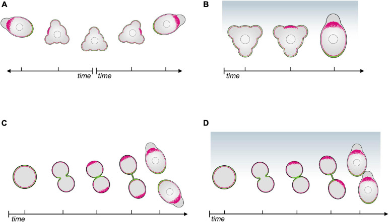
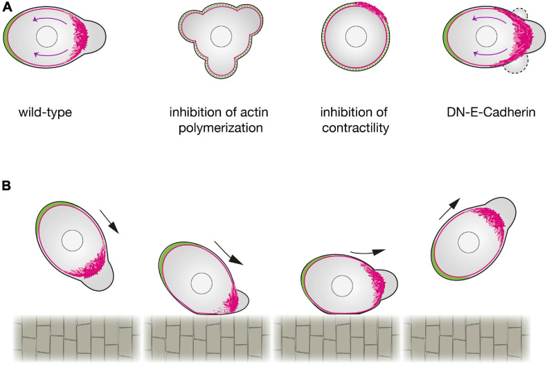
Similar to many other organisms, zebrafish primordial germ cells (PGCs) are specified at a location distinct from that of gonadal somatic cells. Guided by chemotactic cues, PGCs migrate through embryonic tissues toward the region where the gonad develops. In this process, PGCs employ a bleb-driven amoeboid migration mode, characterized by low adhesion and high actomyosin contractility, a strategy used by other migrating cells, such as leukocytes and certain types of cancer cells. The mechanisms underlying the motility and the directed migration of PGCs should be robust to ensure arrival at the target, thereby contributing to the fertility of the organism. These features make PGCs an excellent model for studying guided single-cell migration in vivo. In this review, we present recent findings regarding the establishment and maintenance of cell polarity that are essential for motility and discuss the mechanisms by which cell polarization and directed migration are controlled by chemical and physical cues.

Keywords: Cxcr4; PGC; amoeboid migration; bleb; cell motility; cell polarity; chemokine; chemotaxis.


Conflict of interest statement

The authors declare that the research was conducted in the absence of any commercial or financial relationships that could be construed as a potential conflict of interest.

Figures

FIGURE 1
FIGURE 1
The polarity establishment cascade in zebrafish primordial germ cells in the absence of and in response to chemoattractant. (A) In the absence of chemoattractant, apolar PGCs initiate polarization by enhancing actin polymerization at a random position around the cell perimeter (red); proteins such as Ezrin and Esyt2a (green) accumulate at the opposite side of the cell, that becomes the cell rear. (B) In the presence of a chemokine gradient (Cxcl12a, blue), the cell front (actin in red) is established in the direction of the chemokine source, followed by the definition of the cell rear (green). (C) Upon cell division in the absence of chemoattractant, the linker molecules (Ezrin and Esyt2a in green) accumulate at the cleavage plane, defining the cell rear first; the cell front (actin in red) then forms at the opposite aspect of the cell. (D) When cell division occurs in the presence of a chemokine gradient (Cxcl12a, blue), the leading edge of both daughter cells form in the direction of the source of the chemoattractant.
FIGURE 2
FIGURE 2
Regulation of zebrafish primordial germ cells polarization. (A) A schematic representation of a wild-type PGC (left) compared with a PGC in which actin polymerization (center left), contractility (center right) or cell-cell adhesion (right) are inhibited. Upon a reduction in actin polymerization (by inhibition of Rac1 activity), PGCs cannot form a cell front and polarity is not established. When contractility is blocked (e.g., by inhibition of ROCK activity), PGCs can generate actin brushes, but the cell rear is not defined. When E-Cadherin mediated cell-cell adhesion is inhibited, the actomyosin retrograde flow is enhanced, expanding the area of the cell front where blebs form. Retrograde actin flow is marked by magenta arrows. (B) The effect of contact with a barrier on PGC polarity. PGCs that make contact with the notochord at intermediate or steep angles exhibit a reflective behavior. A reduction in the actin brushes concentration at the side of the cell interacting with the barrier is observed, while the unaffected actin brushes at the opposite side define the new front that directs the migration away from the barrier.

References

    1. Babich A., Li S., O’Connor R. S., Milone M. C., Freedman B. D., Burkhardt J. K. (2012). F-actin polymerization and retrograde flow drive sustained PLCγ1 signaling during T cell activation. J. Cell Biol. 197 775–787. 10.1083/jcb.201201018 - DOI - PMC - PubMed
    1. Bendel-Stenzel M. R., Gomperts M., Anderson R., Heasman J., Wylie C. (2000). The role of cadherins during primordial germ cell migration and early gonad formation in the mouse. Mechan. Develo. 91 143–152. 10.1016/S0925-4773(99)00287-7 - DOI - PubMed
    1. Bisel B., Wang Y., Wei J.-H., Xiang Y., Tang D., Miron-Mendoza M., et al. (2008). ERK regulates Golgi and centrosome orientation towards the leading edge through GRASP65. J. Cell Biol. 182 837–843. 10.1083/jcb.200805045 - DOI - PMC - PubMed
    1. Blaser H., Eisenbeiss S., Neumann M., Reichman-Fried M., Thisse B., Thisse C., et al. (2005). Transition from non-motile behaviour to directed migration during early PGC development in zebrafish. J. Cell Sci. 118 4027–4038. 10.1242/jcs.02522 - DOI - PubMed
    1. Blaser H., Reichman-Fried M., Castanon I., Dumstrei K., Marlow F. L., Kawakami K., et al. (2006). Migration of Zebrafish Primordial Germ Cells: A Role for Myosin Contraction and Cytoplasmic Flow. Devel. Cell 11 613–627. 10.1016/j.devcel.2006.09.023 - DOI - PubMed

LinkOut - more resources