Review
. 2021 Jun 30;3(3):129-133.
doi: 10.35772/ghm.2021.01076.

Effects of COVID-19 vaccination on FDG-PET/CT imaging: A literature review

Affiliations
Review

Effects of COVID-19 vaccination on FDG-PET/CT imaging: A literature review

Ryogo Minamimoto et al. Glob Health Med. .

Abstract

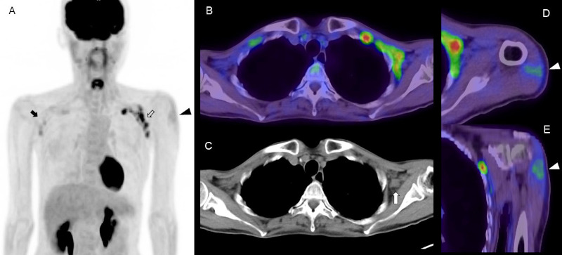
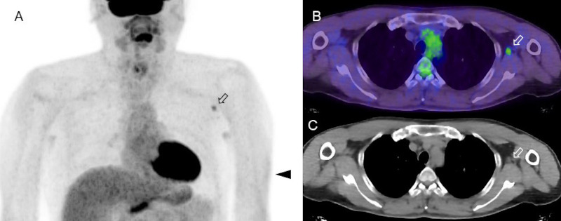
COVID-19 vaccination using mRNA technology began at the end of 2020 in several countries, approximately 9 months after the WHO declared the new coronavirus a pandemic, and began in Japan at the end of February 2021. Several studies have reported FDG avidity in enlarged axillary lymph nodes as a specific feature of FDG-PET/CT imaging after COVID-19 vaccination. A major concern is that this finding could lead to a misdiagnosis in patients with various types of malignancy. We review the impact of COVID-19 vaccination on the management of patients scheduled for FDG-PET/CT in the setting of nationwide mass vaccination.

Keywords: COVID-19; FDG; PET/CT; vaccine.


Conflict of interest statement

The authors have no conflicts of interest to disclose.

Figures

Figure 1.
Figure 1.
FDG PET/CT images in a woman in her 50s with abdominal malignancy at 4 days after COVID-19 vaccination. (A) MIP image, (B) axial fusion image, (C) CT image, (D) axial fusion image of deltoid muscle, (E) coronal fusion image of deltoid muscle. Increased FDG uptake is seen in the left axilla with supraclavicular lymphadenopathy (open arrow). There is slight FDG uptake in the right axillary and supraclavicular areas (arrow). The CT image shows enlarged lymph nodes with loss of fatty hilum in the left axillary and supraclavicular areas (white arrow). Intense FDG uptake in the left deltoid muscle indicates the site of COVID-19 vaccine injection (arrowhead). FDG uptake in the spleen was almost equal to that in the liver, which might have been caused by the vaccination.
Figure 2.
Figure 2.
FDG-PET/CT images in a subject who received an influenza vaccination 4 days before the imaging examination. (A) MIP image, (B) axial fusion image, (C) CT image. Focal FDG uptake is seen in an axillary lymph node, which was slightly enlarged on CT image. Slight FDG uptake in the left arm indicates the site of COVID-19 vaccine injection at subcutaneous tissue (arrowhead). There are slight and symmetrical FDG uptake in the bilateral axillary area due to non-specific uptake for sweat glands.

References

    1. Mitsuya H, Kokudo N. Sustaining containment of COVID-19: global sharing for pandemic response. Glob Health Med. 2020; 2:53-55. - PMC - PubMed
    1. JOHNS HOPKINS CORONAVIRUS RESOURCE CENTER. https://coronavirus.jhu.edu/map.html (last updated at 6/10/2021). (accessed June 21, 2021).
    1. Douglas D, Richman. COVID-19 v a c c ines : implementation, limitations and opportunities. Glob Health Med. 2021; 3:1-5 - PMC - PubMed
    1. Amit S, Regev-Yochay G, Afek A, Kreiss Y, Leshem E. Early rate reductions of SARS-CoV-2 infection and COVID-19 in BNT162b2 vaccine recipients. Lancet. 2021; 397:875-877. - PMC - PubMed
    1. Le TT, Andreadakis Z, Kumar A, Gómez Román R, Tollefsen S, Saville M, Mayhew S. The COVID-19 vaccine development landscape. Nat Rev Drug Discov; 2020: 19:305-306. - PubMed

LinkOut - more resources