Review
. 2021 Jul 27;15(7):11066-11086.
doi: 10.1021/acsnano.1c03087. Epub 2021 Jul 12.

Piezoelectric Nanomaterials Activated by Ultrasound: The Pathway from Discovery to Future Clinical Adoption

Affiliations
Review

Piezoelectric Nanomaterials Activated by Ultrasound: The Pathway from Discovery to Future Clinical Adoption

Andrea Cafarelli et al. ACS Nano. .

Abstract

Electrical stimulation has shown great promise in biomedical applications, such as regenerative medicine, neuromodulation, and cancer treatment. Yet, the use of electrical end effectors such as electrodes requires connectors and batteries, which dramatically hamper the translation of electrical stimulation technologies in several scenarios. Piezoelectric nanomaterials can overcome the limitations of current electrical stimulation procedures as they can be wirelessly activated by external energy sources such as ultrasound. Wireless electrical stimulation mediated by piezoelectric nanoarchitectures constitutes an innovative paradigm enabling the induction of electrical cues within the body in a localized, wireless, and minimally invasive fashion. In this review, we highlight the fundamental mechanisms of acoustically mediated piezoelectric stimulation and its applications in the biomedical area. Yet, the adoption of this technology in a clinical practice is in its infancy, as several open issues, such as piezoelectric properties measurement, control of the ultrasound dose in vitro, modeling and measurement of the piezo effects, knowledge on the triggered bioeffects, therapy targeting, biocompatibility studies, and control of the ultrasound dose delivered in vivo, must be addressed. This article explores the current open challenges in piezoelectric stimulation and proposes strategies that may guide future research efforts in this field toward the translation of this technology to the clinical scene.

Keywords: cancer treatment; electric stimuli; mechanoelectrical transduction; neuromodulation; piezoelectric effect; piezoelectric nanomaterials; regenerative medicine; ultrasound.


Conflict of interest statement

The authors declare no competing financial interest.

Figures
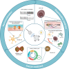
Figure 1
Figure 1
Scheme of the “US-activated piezoelectric nanoparticle stimulation” paradigm and the main research domains in which this paradigm is nowadays explored. US waves can interact with piezoelectric nanoparticles, generating a localized electrical stimulation used for neuromodulation, regeneration, or cancer treatment purposes.
Figure 2
Figure 2
(a) Unit cells of characteristic piezoelectric materials. (i) ZnO: inorganic (nonferroelectric) material with a wurtzite structure. Image adapted with permission from ref (53). Copyright 2009 Elsevier. (ii) BaTiO3: inorganic (ferroelectric) ceramic material with a perovskite structure. The piezoelectric coefficients are defined with respect to the applied stress direction. Image adapted with permission from ref (54). Copyright 2002 John Wiley and Sons. (iii) PVDF: synthetic organic polymer exhibiting high piezoelectricity in the β structure. Image adapted with permission from ref (7). Copyright 2019 John Wiley and Sons. (b) Typical features of a pulsed ultrasound wave at a specific frequency (f): period (T = 1/f), pulse period time (Ton), delay time (Toff), peak of positive pressure (PpP), and peak of negative pressure (PnP). These parameters also allow calculating the pulse repetition period (PRP = Ton + Toff), duty cycle (DC = Ton/PRP), and burst rate (BR = 1/PRP); intensity (I) is derived by dividing the square of the pressure (PUS) by the density of the medium (ρ) and the traveling wave speed (c).
Figure 3
Figure 3
Representative images of the main findings achieved by applying the “US-activated piezoelectric nanoparticle stimulation” paradigm to electrically excitable cells. (a) Barium titanate nanoparticles (BTNPs) internalized within SH-SY5Y-derived neurons (i); when ultrasound (US) is applied, a significantly higher calcium flux is detected (ii). Images reproduced from ref (55). Copyright 2015 American Chemical Society. (b) Enhancement of the firing rate in neurons provided with BTNPs and stimulated with US. Images reproduced with permission from ref (83). Copyright 2020 Springer Nature. (c) Depiction of the experimental procedure (i) and photos showing changes in the spontaneous coiling behavior on a zebrafish embryo induced by BTNPs and US (ii). Images adapted with permission from ref (56). Copyright 2020 John Wiley and Sons. (d) Enhancement of neurite elongation in PC12 cells internalizing BNNTs and stimulated with US. Images reproduced from ref (58). Copyright 2010 American Chemical Society. (e) US+BTNPs enhances Ca2+ transients in SH-SY5Y cells. Image reproduced with permission from ref (63). Copyright 2016 John Wiley and Sons. (f) US stimulation of S. platentis with BTNPs (i) triggers different intracellular pathways affecting PC12 cell differentiation (ii) and mediating neurite outgrowth (iii). Images reproduced with permission from ref (73). Copyright 2020 John Wiley and Sons. (g) C2C12 cells internalizing BNNTs and stimulated with US receive a boost to form multinucleated myotubes featured by a higher fusion index (index of skeletal muscle tissue maturity). Images reprinted with permission under a Creative Commons Attribution License from ref (59). Copyright 2013 Ricotti et al.
Figure 4
Figure 4
Representative images of the main findings achieved by applying the “piezonanoparticles + US” paradigm to non-electrically excitable cells. (a) BNNTs in combination with US up-regulate calcium production and osteopontin expression in osteoblasts. Image reproduced with permission from ref (60). Copyright 2013 IOP Publishing. (b) Ag-conjugated barium titanate nanoparticles (BTNPs) (i) stimulated with US enhance the proliferation of osteosarcoma-derived cells (ii). Images adapted with permission from ref (72). Copyright 2020 Elsevier. (c) F-actin overexpression in dermal fibroblasts triggered by US + BNNTs. Images reproduced with permission from ref (61). Copyright 2014 Springer Nature. (d) US enhances proliferation of fibroblasts in the presence of BTNPs, embedded in a scaffold. Images reproduced with permission from ref (64). Copyright 2017 Elsevier. (e) Combination of US and BTNPs down-regulates the Ki-67 proliferative marker in breast cancer cells (i), induces the arrest of the cell cycle in G0/G1 phases (ii), and increases the intracellular concentration of calcium (iii). Images reprinted with permission under a Creative Commons CC BY License from ref (66). Copyright 2018 Springer Nature. (f) US-induced piezoelectric treatment induces cell apoptosis and decreases proliferation in glioblastoma cells. Images adapted with permission from ref (69). Copyright 2019 Elsevier.
Figure 5
Figure 5
Scheme of the key aspects to be addressed to foster clinical acceptance of the “US-activated piezoelectric nanoparticle stimulation” paradigm. The identified route includes the following substeps. (a) Piezoelectric properties measurement: accurate quantification of the piezoelectric coefficients plays a crucial role in the experimental design phase and material selection. (b) Control of the US dose released in vitro: the use of dose-controlled stimulation systems enables a precise correlation between the effective US dose and the biological findings. (c) Quantification of US-mediated piezo effect: the interaction between US waves and piezoelectric particles needs to be further explored for a better understanding of the underlying phenomenon. (d) Knowledge of related bioeffects: the activated cellular mechanisms need to be elucidated more in-depth. (e) Therapy targeting: new approaches for enhancing the spatial localization of the therapy delivery result crucial for a targeted in vivo use. (f) Biocompatibility studies: careful analyses about nanoparticles biosafety have to be accomplished before their use in the clinics. (g) Control of the US dose released in vivo: US must be correctly tuned in order to reach the target in vivo with the desired dose.

References

    1. Balint R.; Cassidy N. J.; Cartmell S. H. Electrical Stimulation: A Novel Tool for Tissue Engineering. Tissue Eng., Part B 2013, 19 (1), 48–57. 10.1089/ten.teb.2012.0183. - DOI - PubMed
    1. Wells P. N. T. Ultrasound Imaging. Phys. Med. Biol. 2006, 51 (13), R83.10.1088/0031-9155/51/13/R06. - DOI - PubMed
    1. Escoffre J.-M., Bouakaz A., Eds. Therapeutic Ultrasound; Advances in Experimental Medicine and Biology; Springer International Publishing: Cham, Switzerland, 2016; Vol. 880.
    1. Hsiao Y.-H.; Kuo S.-J.; Tsai H.-D.; Chou M.-C.; Yeh G.-P. Clinical Application of High-Intensity Focused Ultrasound in Cancer Therapy. J. Cancer 2016, 7 (3), 225–231. 10.7150/jca.13906. - DOI - PMC - PubMed
    1. Jiang X.; Savchenko O.; Li Y.; Qi S.; Yang T.; Zhang W.; Chen J. A Review of Low-Intensity Pulsed Ultrasound for Therapeutic Applications. IEEE Trans. Biomed. Eng. 2019, 66 (10), 2704–2718. 10.1109/TBME.2018.2889669. - DOI - PubMed

Publication types