. 2021 Oct 15;204(8):933-942.
doi: 10.1164/rccm.202101-0122OC.

Addition of 5% CO2 to Inspiratory Gas Prevents Lung Injury in an Experimental Model of Pulmonary Artery Ligation

Affiliations

Addition of 5% CO2 to Inspiratory Gas Prevents Lung Injury in an Experimental Model of Pulmonary Artery Ligation

Ines Marongiu et al. Am J Respir Crit Care Med. .

Abstract

Rationale: Unilateral ligation of the pulmonary artery may induce lung injury through multiple mechanisms, which might be dampened by inhaled CO2. Objectives: This study aims to characterize bilateral lung injury owing to unilateral ligation of the pulmonary artery in healthy swine undergoing controlled mechanical ventilation and its prevention by 5% CO2 inhalation and to investigate relevant pathophysiological mechanisms. Methods: Sixteen healthy pigs were allocated to surgical ligation of the left pulmonary artery (ligation group), seven to surgical ligation of the left pulmonary artery and inhalation of 5% CO2 (ligation + FiCO2 5%), and six to no intervention (no ligation). Then, all animals received mechanical ventilation with Vt 10 ml/kg, positive end-expiratory pressure 5 cm H2O, respiratory rate 25 breaths/min, and FiO2 50% (±FiCO2 5%) for 48 hours or until development of severe lung injury. Measurements and Main Results: Histological, physiological, and quantitative computed tomography scan data were compared between groups to characterize lung injury. Electrical impedance tomography and immunohistochemistry analysis were performed in a subset of animals to explore mechanisms of injury. Animals from the ligation group developed bilateral lung injury as assessed by significantly higher histological score, larger increase in lung weight, poorer oxygenation, and worse respiratory mechanics compared with the ligation + FiCO2 5% group. In the ligation group, the right lung received a larger fraction of Vt and inflammation was more represented, whereas CO2 dampened both processes. Conclusions: Mechanical ventilation induces bilateral lung injury within 48 hours in healthy pigs undergoing left pulmonary artery ligation. Inhalation of 5% CO2 prevents injury, likely through decreased stress to the right lung and antiinflammatory effects.

Keywords: CO2 inhalation; VILI; pulmonary perfusion; therapeutic hypercapnia.


Figures

Figure 1.
Figure 1.
Severity of histological, computed tomographic scan, and physiological alterations at the end of the experiment. (A) The histological score of lungs from each study group. The score had 10 components that were ranked between 0 and 3 and summed within apical, medial, and basal samples from each lung. Then, scores from the six samples were averaged to obtain the total lung histological score for each animal (range 0–30). (B) Quantitative computed tomographic scan analysis showing change in lung weights from the baseline (after instrumentation and before surgical ligation of the left pulmonary artery) to the end of the experiment (48 hours or at development of severe lung injury) (Δlung weight) in each study group. (C) Proportion of nonaerated lung tissue in each study group. (D and E) Compliance of the respiratory system (rs) (D) and PaO2/FiO2 (E) at the end of the experiment in each study group. Data are expressed as scatter dot plot with mean ± SEM. Statistical analysis was performed by one-way ANOVA followed by post hoc Holm-Sidak test (A, C, D, and E) or Kruskal-Wallis test (B). P values in the graph refer to ANOVA/Kruskal-Wallis P value. *P < 0.05 and ***P < 0.001, ligation + FiCO2 5% versus ligation group. ^P < 0.05, ^^P < 0.01, and ^^^P < 0.01, no-ligation versus ligation group.
Figure 2.
Figure 2.
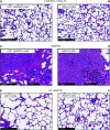
Microscopic appearance of the lungs at the end of the experiment. (A–D) Representative microphotographs of the left ligated and right nonligated lungs from the ligation + FiCO2 5% group (A and B) and ligation group (C and D). (C) The left lung of the ligation group shows a marked inflammatory infiltrate composed primarily of lymphocytes and macrophages, with vascular congestion and hemorrhage. (D) The right lung of the ligation group shows a patchy acute inflammatory infiltrate composed primarily of neutrophils (hematoxylin and eosin [H&E]). Notably, the lungs of the ligation + FiCO2 5% group (A and B) showed no inflammatory changes (H&E). (E and F) In the lower panels, representative microphotographs of the lungs from the no ligation group with no significant histological alterations (H&E). Scale bars: main panels, 500 μm; insets, 100 μm.
Figure 3.
Figure 3.
Ventilation distribution and regional respiratory system compliance along the experiments. (A) The ratio between Vt distending the right and the left lung along the study time points shows significant imbalance in the ligation group, which was prevented by inhalation of CO2. (B and C) Respiratory system compliance for left (B) and right (C) respiratory hemisystem. In the ligation group, left compliance was lower than the ligation + FiCO2 group and remained stable over time, whereas in the right side of the ligation group, it declined along the study; FiCO2 maintained both compliances stable. Data are expressed as mean ± SEM. Statistical analysis was performed using generalized estimating equation models to account for repeated measures in time (longitudinal data); the model included group and time as main independent factors and group-by-time interaction. *P < 0.05 and **P < 0.01, ligation + FiCO2 5% versus ligation group. Compliance rsL = respiratory system compliance for left respiratory hemisystem; Compliance rsR = respiratory system compliance for right respiratory hemisystem; VtRL/VtLL = the ratio between Vt distending the right and the left lung.
Figure 4.
Figure 4.
Characterization of the lung immune cell infiltrates in the different groups by immunohistochemistry. (A and C) Representative image of the MPO (myeloperoxidase)-positive (A) or IBA-1 (ionized calcium-binding adaptor 1)–positive (C) infiltrates in the lungs of pigs from the three different groups of treatment with the corresponding digital quantification (mask). Images show representative samples from the most affected side. Scale bars, 100 μm. (B and D) Quantification of the MPO-positive (B) or IBA-1–positive (D) cells in the different groups. Each circle is a sample (n = 8 per group); bars, mean ± SEM. (B) *q = 0.010, no ligation versus ligation; **q = 0.008, ligation versus ligation + FiCO2 5%. (D) ^^q = 0.001, no ligation versus ligation + FiCO2 5%; *q = 0.014, no ligation versus ligation + FiCO2 5%. P values from Kruskal-Wallis tests are reported in the graphs; q values are false discovery rate–adjusted P values from Dunn’s post hoc test according to the Benjamini, Krieger, and Yekutieli method.
Figure 5.
Figure 5.
Differences between left ligated and right nonligated lung in the ligation group. (A) Histological score of each lung in the ligation group at the end of the experiment. (B) Quantitative computed tomographic scan analysis showing change in lung weight of each lung in the ligation group from the baseline (after instrumentation and before surgical ligation of the left pulmonary artery) to the end of the experiment (48 h or at development of severe lung injury) (Δlung weight). (C) Proportion of nonaerated lung tissue of each lung in the ligation group at the end of the experiment. Data are expressed as scatter dot plot with mean ± SEM. Statistical analysis was performed by a paired t test, and P values are reported in the graph.
Figure 6.
Figure 6.
Pathophysiological mechanisms inducing bilateral lung injury and protective effects of inhaled CO2. Left pulmonary artery ligation induces bilateral lung injury through multiple processes (black pathway). The addition of 5% CO2 to inspiratory gases guarantees alveolar normocapnia and exerts antiinflammatory properties, blocking critical processes and ultimately preventing injury (green pathways).

Comment in

References

    1. Edmunds LH, Jr, Holm JC. Effect of inhaled CO2 on hemorrhagic consolidation due to unilateral pulmonary arterial ligation. J Appl Physiol. 1969;26:710–715. - PubMed
    1. Kolobow T, Spragg RG, Pierce JE. Massive pulmonary infarction during total cardiopulmonary bypass in unanesthetized spontaneously breathing lambs. Int J Artif Organs. 1981;4:76–81. - PubMed
    1. Greene R, Zapol WM, Snider MT, Reid L, Snow R, O’Connell RS, et al. Early bedside detection of pulmonary vascular occlusion during acute respiratory failure. Am Rev Respir Dis. 1981;124:593–601. - PubMed
    1. Nuckton TJ, Alonso JA, Kallet RH, Daniel BM, Pittet JF, Eisner MD, et al. Pulmonary dead-space fraction as a risk factor for death in the acute respiratory distress syndrome. N Engl J Med. 2002;346:1281–1286. - PubMed
    1. Sinha P, Calfee CS, Beitler JR, Soni N, Ho K, Matthay MA, et al. Physiologic analysis and clinical performance of the ventilatory ratio in acute respiratory distress syndrome. Am J Respir Crit Care Med. 2019;199:333–341. - PMC - PubMed

Publication types