. 2021 Aug 21;42(32):3078-3090.
doi: 10.1093/eurheartj/ehab431.

PCSK9 deficiency rewires heart metabolism and drives heart failure with preserved ejection fraction

Affiliations

PCSK9 deficiency rewires heart metabolism and drives heart failure with preserved ejection fraction

Lorenzo Da Dalt et al. Eur Heart J. .

Abstract

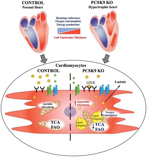
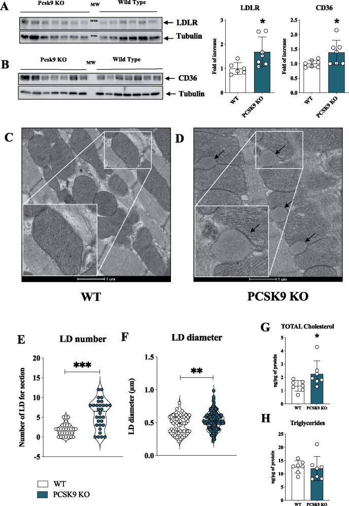
Aims: PCSK9 is secreted into the circulation, mainly by the liver, and interacts with low-density lipoprotein receptor (LDLR) homologous and non-homologous receptors, including CD36, thus favouring their intracellular degradation. As PCSK9 deficiency increases the expression of lipids and lipoprotein receptors, thus contributing to cellular lipid accumulation, we investigated whether this could affect heart metabolism and function.

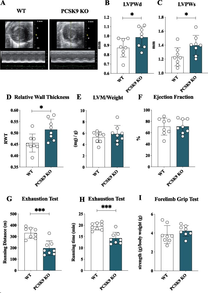
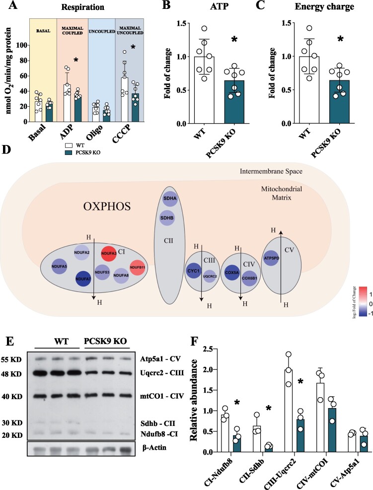
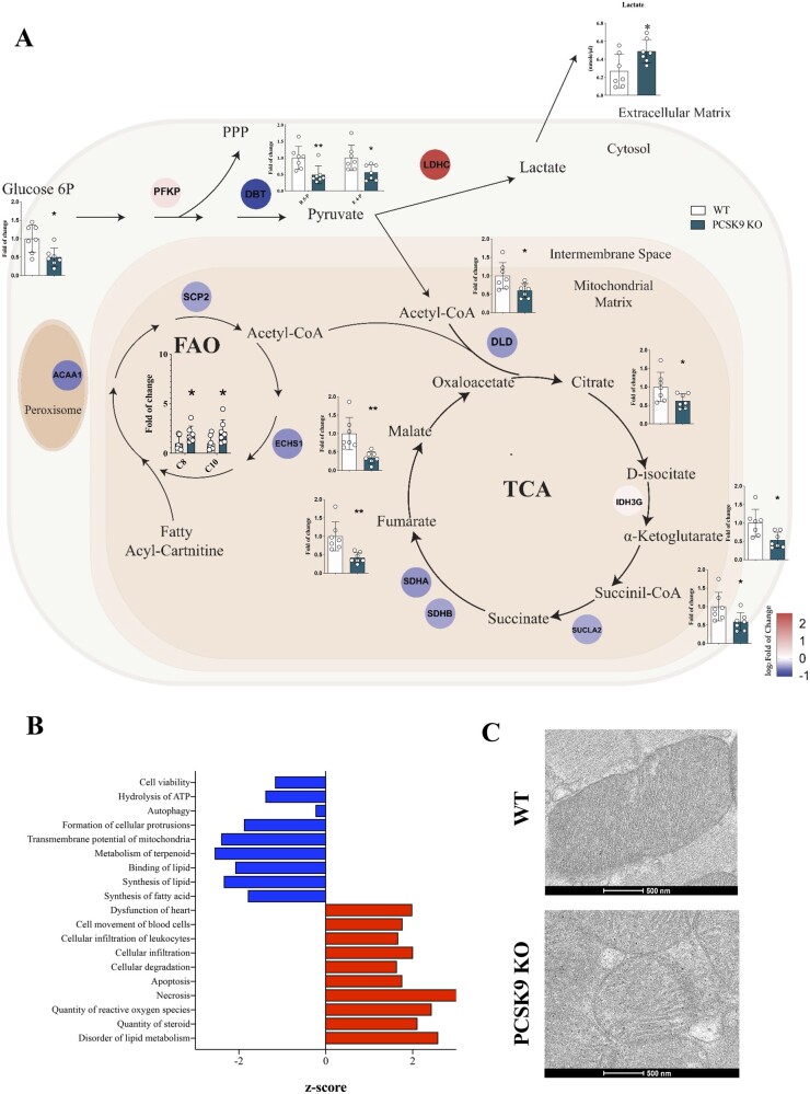
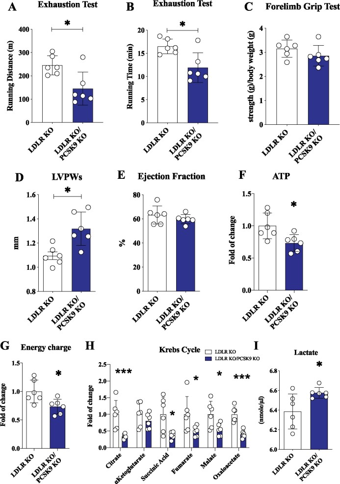
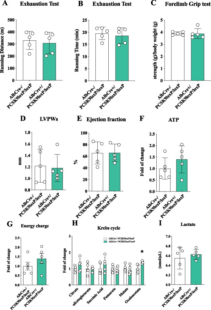
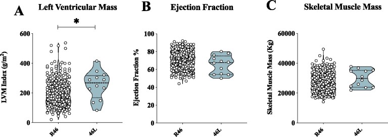
Methods and results: Wild-type (WT), Pcsk9 KO, Liver conditional Pcsk9 KO and Pcsk9/Ldlr double KO male mice were fed for 20 weeks with a standard fat diet and then exercise resistance, muscle strength, and heart characteristics were evaluated. Pcsk9 KO presented reduced running resistance coupled to echocardiographic abnormalities suggestive of heart failure with preserved ejection fraction (HFpEF). Heart mitochondrial activity, following maximal coupled and uncoupled respiration, was reduced in Pcsk9 KO mice compared to WT mice and was coupled to major changes in cardiac metabolism together with increased expression of LDLR and CD36 and with lipid accumulation. A similar phenotype was observed in Pcsk9/Ldlr DKO, thus excluding a contribution for LDLR to cardiac impairment observed in Pcsk9 KO mice. Heart function profiling of the liver selective Pcsk9 KO model further excluded the involvement of circulating PCSK9 in the development of HFpEF, pointing to a possible role locally produced PCSK9. Concordantly, carriers of the R46L loss-of-function variant for PCSK9 presented increased left ventricular mass but similar ejection fraction compared to matched control subjects.

Conclusion: PCSK9 deficiency impacts cardiac lipid metabolism in an LDLR independent manner and contributes to the development of HFpEF.

Keywords: Cholesterol; HFpEF; Heart; LDLR; PCSK9.


Figures

None
Impact of Pcsk9 deficiency on cardiac function and mitochondrial metabolism. Pcsk9 deficiency is associated with increased heart left ventricular thickness and reduced running performance independently of skeletal muscle alterations. Electron microscopy analysis showed increased cardiac accumulation of lipid droplets associated with a reduced density of mitochondrial cristae; this proffunctionally translated into impaired oxidative phosphorylation and mitochondrial metabolism in PCSK9 KO hearts.
Figure 1
Figure 1
Pcsk9 deficiency is associated with heart failure with preserved ejection fraction. (A) Representative image of echocardiographic analysis for wild-type and Pcsk9 KO mice. (B, C) Left ventricular posterior wall thickness during systole (P = 0.049) and diastole (P = 0.03). (D–F) relative wall thickness (RWT) (P = 0.02), the left ventricular mass/weight (P = 0.37) and ejection fraction (%) (P = 0.95) are shown. (G, H) Running endurance following exhaustion test for wild-type and Pcsk9 KO mice is presented as running distance (P = 0.0003) and running time (P = 0.0003). (I) Results from the forelimb grip test are presented (P = 0.33). Data are shown as mean ± SD; n = 8 mice per group. Non-parametric t-test was used to compare each group. *P < 0.05 and ***<0.001.
Figure 2
Figure 2
Pcsk9 deficiency is associated with mitochondrial dysfunction. (A) The oxygen consumption rate was investigated in the heart of Pcsk9 KO mice and oxygen consumption was measured at basal, maximal coupled, uncoupled and maximal uncoupled conditions (Basal, P = 0.39; ADP, P = 0.04; Oligo, P = 0.24; CCCP, P = 0.04). Data are shown as mean ± SD; n = 7 mice per group. (B and C) Adenosine triphosphate (P = 0.01) quantification and energy charge (P = 0.01) of the heart. Data are shown as fold of change ± SD; n = 7 mice per group. (D) Relevant proteins of ETC are displayed. (E) Representative image of western blot of ETC complexes on heart lysate is showed. (F) Proteins quantification is normalized to beta-actin expression (CI, P = 0.01; CII, P = 0.02; CIII, P = 0.008; CIV, P = 0.08, CV, P = 0.59). Data are shown as mean ± SD; n = 3 mice per group. Non-parametric t-test was used to compare each group (*P < 0.05).
Figure 3
Figure 3
Metabolic profile of failing Pcsk9 KO heart. (A) Results from combined metabolomic, proteomic and lipidomic profile in the heart of Pcsk9 KO mice compared to wild-type mice are shown. Metabolites that are significantly modulated (belonging to glycolysis, pentose phosphate pathway, Krebs cycle, or carnitines for beta-oxidation) (G6-P, P = 0.012; R5-P, P = 0.01; E4-P, P = 0.02; acetyl-CoA, P = 0.02; citrate, P = 0.04; α-ketoglutarate, P = 0.01; succinyl-CoA, P = 0.03; fumarate, P = 0.003; malate, P = 0.003; C8, P = 0.048; C10, P = 0.048) are shown in bar graph as fold of change ± SD; n = 7 mice per group. Proteins that were significantly modulated following proteomics analysis are shown as coloured dots. Plasma levels of lactate are shown (P = 0.02). Data are shown as mean ± SD; n = 7 per group. (B) Functional pathway analysis of proteomics data is presented. Hierarchical clustering is based on Pearson’s correlation and heatmap showing relative protein expression values (z-score-transformed LFQ protein intensities) of n=65 proteins corresponding to ETC mitochondrial complexes in GO analysis (FDR < 0.05). Non-parametric t-test was used to compare each group. (C) Representative photomicrographs of myocardium mitochondria by transmission electron microscopy are shown. (*P < 0.05 and **<0.01)
Figure 4
Figure 4
PCSK9 deficiency results in increased lipids and lipoprotein receptor expression coupled with cholesterol accumulation and the increase of lipid droplets in the heart. (A) Representative image and quantification of immunoblotting analysis for LDLR in cardiac tissue from Pcsk9 KO and wild-type mice (P = 0.03). Data are shown as mean ± SD; n = 6 mice for the wild-type group and n = 7 mice for the Pcsk9 KO group. (B) Representative image and quantification of immunoblotting analysis for CD36 in cardiac tissue from Pcsk9 KO and wild-type mice (P = 0.04). Data are shown as mean ± SD; n = 7 mice for group. (C and D) Representative photomicrographs of myocardium by transmission electron microscopy in wild-type and Pcsk9 KO mice are shown. Black arrows indicate lipid droplets. The inset panels show a magnification of wild-type mice (C) and of Pcsk9 KO mice (D). Bars 1 µm. (E and F) Lipid droplets number (P < 0.0001) and diameter (P = 0.008) obtained from transmission electron microscopy analysis are shown as violin plot. (G) Intracardiac total cholesterol (P = 0.04) and (E) triglycerides levels (P = 0.77) are shown. Data are presented as mean ± SD; n = 7 mice for group. Non-parametric t-test was used to compare each group (*P < 0.05, **P < 0.01 and ***<0.001).
Figure 5
Figure 5
PCSK9 effect on cardiac function is not dependent on the LDLR. (A and B) Running endurance on exhaustion test of Ldlr KO and Pcsk9/Ldlr DKO mice is displayed as running distance (P = 0.01) and running time (P = 0.01). (C) Results from the forelimb grip test are displayed (P = 0.23). (D and E) Left ventricular posterior wall thickness during systole (P = 0.01) and ejection fraction (P = 0.33) are shown. (F and G) Adenosine triphosphate quantification (P = 0.02) and energy charge (P = 0.02) of the heart. Data are presented as mean ± SD. (H) Metabolites of the Krebs cycle are shown (citrate, P = 0.003; α-ketoglutarate, P = 0.29; succinyl-CoA, P = 0.02; fumarate, P = 0.05; malate, P = 0.03; oxaloacetate, P = 0.0002) as fold of change ± SD. (I) Plasma levels of lactate (P = 0.03) are shown as mean ± SD. n = 6 mice per group. Non-parametric t-test was used to compare each group (*P < 0.05 and ***<0.001).
Figure 6
Figure 6
Circulating PCSK9 does not impact cardiac metabolism and heart structure. (A and B) Running endurance on exhaustion test of AlbCre−/Pcsk9LoxP/LoxP and AlbCre+/Pcsk9LoxP/LoxP mice is displayed as running distance (P = 0.68) and running time (P = 0.63). (C) Results from the forelimb grip test are displayed (P = 0.98). (D and E) Left ventricular posterior wall thickness during systole (P = 0.83) and ejection fraction (P = 0.98) are shown. (F and G) Adenosine triphosphate quantification (P = 0.27) and energy charge (P = 0.627) of the heart are presented. (H) Metabolites of the Krebs cycle (citrate, P = 0.26; α-ketoglutarate, P = 0.91; succinyl-CoA, P = 0.33; fumarate, P = 0.57; malate, P = 0.63; oxaloacetate, P = 0.009) are shown as fold of change ± SD. (I) Plasma levels of lactate (P = 0.50) are shown. Data are shown as mean ± SD; n = 5 mice per group. Non-parametric t-test was used to compare each group (*P < 0.05).
Figure 7
Figure 7
PCSK9 46L LOF individuals present heart failure with preserved ejection fraction. (A and B) Left ventricular mass index (g/m2) (P = 0.01) and ejection fraction (P = 0.10) of PCSK9 R46 and 46L carriers are shown. (C) Leg and arm skeletal muscle mass (quantified by Dual X-Rays Absorbimetry, following Hansen’s formula) (P = 0.26) is shown (R46, n = 516; 46L, n = 12). Non-parametric t-test was used to compare each group. (*P < 0.05).

Comment in

  • Heart to heart with PCSK9.
    Chemello K, Jaafar AK, Lambert G. Chemello K, et al. Eur Heart J. 2021 Aug 21;42(32):3091-3093. doi: 10.1093/eurheartj/ehab480. Eur Heart J. 2021. PMID: 34324660 No abstract available.

References

    1. Stanley WC, Recchia FA, Lopaschuk GD. Myocardial substrate metabolism in the normal and failing heart. Physiol Rev 2005;85:1093–1129. - PubMed
    1. Pulinilkunnil T, Rodrigues B. Cardiac lipoprotein lipase: metabolic basis for diabetic heart disease. Cardiovasc Res 2006;69:329–340. - PubMed
    1. van der Vusse GJ, van Bilsen M, Glatz JF. Cardiac fatty acid uptake and transport in health and disease. Cardiovasc Res 2000;45:279–293. - PubMed
    1. Bharadwaj KG, Hiyama Y, Hu Y, Huggins LA, Ramakrishnan R, Abumrad NA, Shulman GI, Blaner WS, Goldberg IJ. Chylomicron- and VLDL-derived lipids enter the heart through different pathways: in vivo evidence for receptor- and non-receptor-mediated fatty acid uptake. J Biol Chem 2010;285:37976–37986. - PMC - PubMed
    1. Goldberg IJ, Trent CM, Schulze PC. Lipid metabolism and toxicity in the heart. Cell Metab 2012;15:805–812. - PMC - PubMed

Publication types