Clinical Trial
. 2021 Jul 12;23(1):72.
doi: 10.1186/s13058-021-01442-7.

Intraoperative fluorescence imaging with aminolevulinic acid detects grossly occult breast cancer: a phase II randomized controlled trial

Affiliations
Clinical Trial

Intraoperative fluorescence imaging with aminolevulinic acid detects grossly occult breast cancer: a phase II randomized controlled trial

Kathryn Ottolino-Perry et al. Breast Cancer Res. .

Abstract

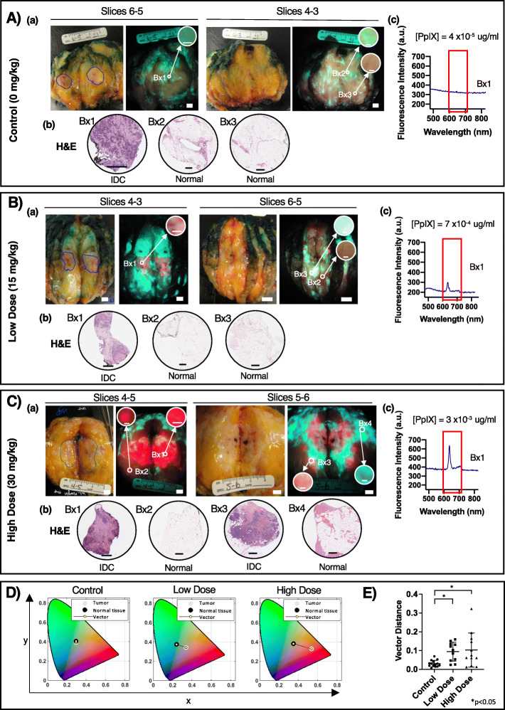
Background: Re-excision due to positive margins following breast-conserving surgery (BCS) negatively affects patient outcomes and healthcare costs. The inability to visualize margin involvement is a significant challenge in BCS. 5-Aminolevulinic acid hydrochloride (5-ALA HCl), a non-fluorescent oral prodrug, causes intracellular accumulation of fluorescent porphyrins in cancer cells. This single-center Phase II randomized controlled trial evaluated the safety, feasibility, and diagnostic accuracy of a prototype handheld fluorescence imaging device plus 5-ALA for intraoperative visualization of invasive breast carcinomas during BCS.

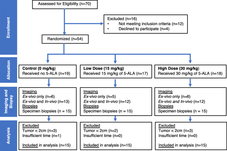
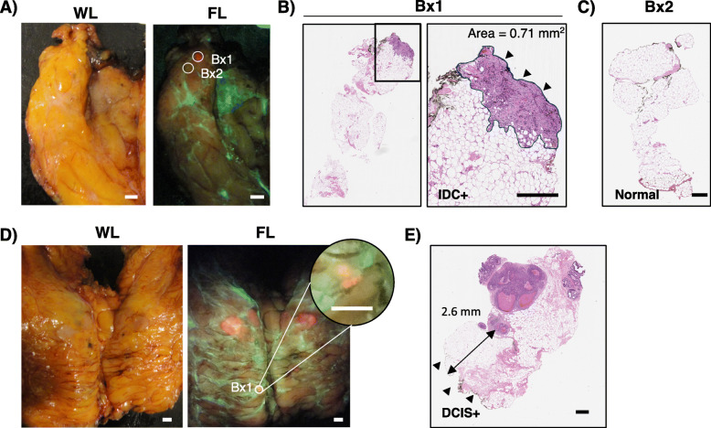
Methods: Fifty-four patients were enrolled and randomized to receive no 5-ALA or oral 5-ALA HCl (15 or 30 mg/kg). Forty-five patients (n = 15/group) were included in the analysis. Fluorescence imaging of the excised surgical specimen was performed, and biopsies were collected from within and outside the clinically demarcated tumor border of the gross specimen for blinded histopathology.

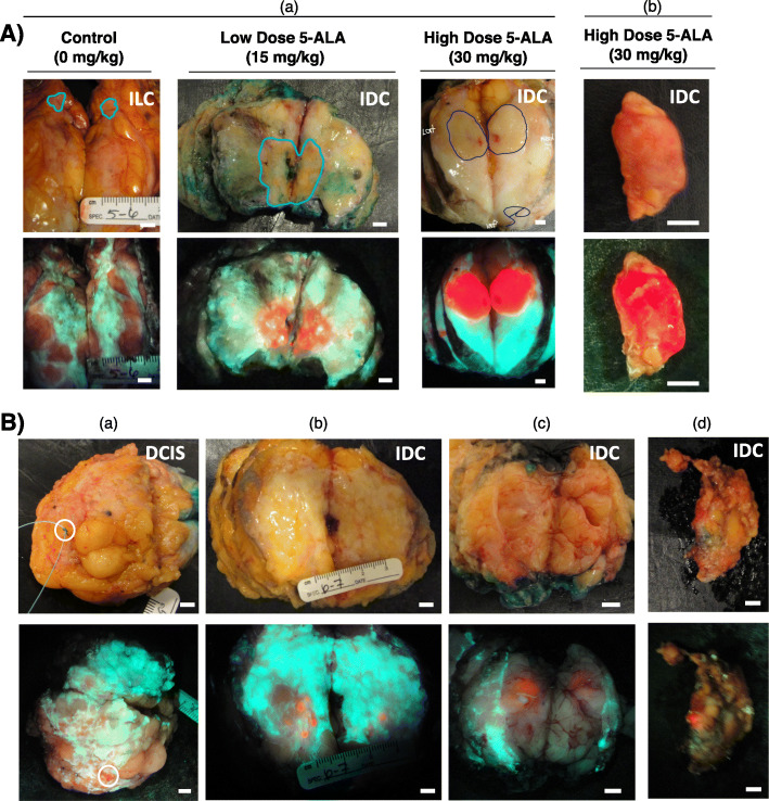
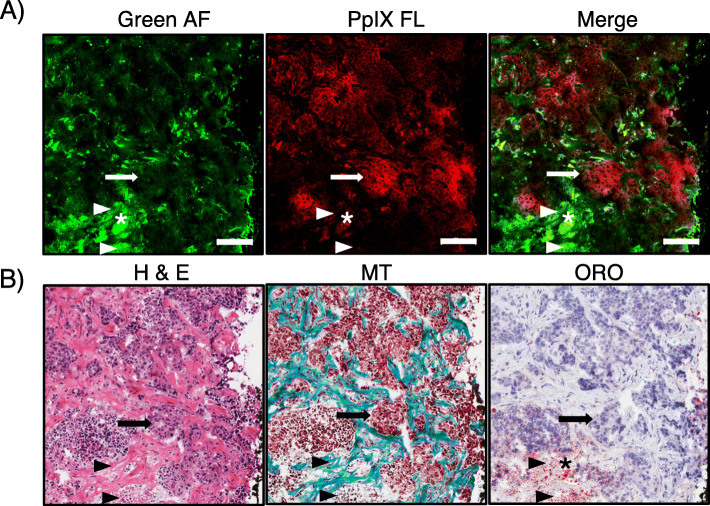
Results: In the absence of 5-ALA, tissue autofluorescence imaging lacked tumor-specific fluorescent contrast. Both 5-ALA doses caused bright red tumor fluorescence, with improved visualization of tumor contrasted against normal tissue autofluorescence. In the 15 mg/kg 5-ALA group, the positive predictive value (PPV) for detecting breast cancer inside and outside the grossly demarcated tumor border was 100.0% and 55.6%, respectively. In the 30 mg/kg 5-ALA group, the PPV was 100.0% and 50.0% inside and outside the demarcated tumor border, respectively. No adverse events were observed, and clinical feasibility of this imaging device-5-ALA combination approach was confirmed.

Conclusions: This is the first known clinical report of visualization of 5-ALA-induced fluorescence in invasive breast carcinoma using a real-time handheld intraoperative fluorescence imaging device.

Trial registration: Clinicaltrials.gov identifier NCT01837225 . Registered 23 April 2013.

Keywords: Aminolevulinic acid; Breast cancer; Breast-conserving surgery; Fluorescence imaging; Handheld intraoperative imaging device; Intraoperative imaging; Margin assessment; Optical imaging.


Conflict of interest statement

RSD, KOP, CG, NTA, WL, AE, and SD are co-inventors on IP licensed to MolecuLight Inc. (Toronto, Canada) and photonamic GmbH & Co. KG (Pinneberg, Germany) commercializing this technology. This study was a University Health Network (UHN) investigator-sponsored RCT (clinicaltrials.gov identifier NCT01837225) prior to IP licensing. RSD is the Founder, Chief Scientific Officer, Board member, and Shareholder of MolecuLight Inc. MolecuLight Inc. did not provide funding or other support, nor were any company staff involved directly or indirectly with this study. RSD conceived of the study and reviewed and edited the final manuscript. He did not recruit nor consent patients, nor collect, nor analyze data resulting from the study. 5-ALA HCl was provided by photonamic GmbH and Co. KG (Pinneberg, Germany) governed by a Drug Transfer Agreement between photonamic and UHN. RSD holds interest in photonamic GmbH & Co. KG’s subsidiary company (SBI ALApharma Canada Inc., Toronto, Canada) developing this technology for commercialization. KOP is employed by SBI ALApharma Canada Inc., but was solely employed by UHN during study data collection, data analysis and manuscript preparation. SBI ALApharma Canada Inc. was not involved directly or indirectly in this study. RSD is governed by a UHN Conflict of Interest Management Plan. All other authors declare that they have no competing interests.

Figures

Fig. 1
Fig. 1
CONSORT diagram. Patients with tumors measuring < 2 cm (greatest dimension) at specimen gross examination were excluded from the analysis because biopsies could not be collected from inside the demarcated tumor border. One case was excluded because of insufficient time to complete data collection prior to initiating formalin fixation of the specimen
Fig. 2
Fig. 2
Fluorescence imaging of 5-ALA-induced PpIX fluorescence in grossly obvious and grossly occult carcinoma. A Representative white light (top row) and fluorescence (bottom row) images of grossly obvious disease in sectioned lumpectomy specimens (a) and a clinically positive sentinel lymph node (c). The pathologist’s assistant (V.S., M.S., F.M.) demarcated tumor border (blue line) identified the grossly obvious tumor in the sectioned specimens. The surgeon (A.M.E., W.L.L.) identified the lymph node as grossly obvious for disease. B Representative white light (top row) and fluorescence (bottom row) images of grossly occult disease at the surface of an excised lumpectomy (a), in grossly sectioned specimens (b, c) and a sentinel lymph node (d) from patients with invasive ductal carcinoma with (a, d) or without (b, c) a DCIS component administered 15 mg/kg (b, c) or 30 mg/kg (a, d) 5-ALA HCl. Images represent tissue that was identified by the surgeon (A.M.E., W.L.L.) (a, d) or pathologist’s assistant (V.S., M.S., F.M.) (b, c) as grossly negative for the presence of cancer. (a) DCIS identified by fluorescence imaging at the lumpectomy margin. (b, c) Invasive carcinoma identified by fluorescence imaging on slices outside the grossly demarcated tumor. (d) Invasive carcinoma macro-metastases identified by fluorescence imaging in an excised sentinel lymph node. Scale bars = 5 mm
Fig. 3
Fig. 3
Detection of grossly occult sub-millimeter red fluorescence tumor foci. A WL and fluorescence images of a slice containing no clinically obvious disease from a patient who received 30 mg/kg 5-ALA HCl. Biopsies were collected in an area of focal red PpIX fluorescence (Bx1) and an adjacent area lacking PpIX fluorescence (Bx2). B H&E-stained longitudinal section of the Bx1 biopsy identified in A, which was determined to contain invasive ductal carcinoma by a blinded pathologist (S.J.D.). The imaged surface of the biopsy is indicated by the arrowheads. The area of tumor near the imaged surface measured 0.71 mm2. C H&E-stained longitudinal section of the Bx2 biopsy identified in A, which was determined to be negative for tumor by a blinded pathologist (S.J.D.). D White light and fluorescence images of slices of a lumpectomy from a patient who received 15 mg/kg 5-ALA HCl. A biopsy (Bx1) was collected from a small area of red fluorescence (inset digitally zoomed). E H&E-stained longitudinal section of the Bx1 identified in D, which was determined to contain DCIS > 2 mm below the imaged surface (arrowheads). Scale bar = 0.5 mm (A, D), 500 μm (B, C, E). WL, white light; FL, fluorescence; Bx, biopsy; IDC, invasive ductal carcinoma; DCIS, ductal carcinoma in situ
Fig. 4
Fig. 4
Cancer cell-specific localization of ALA-induced PpIX fluorescence. Representative A fluorescence microscopy and B corresponding histological images (bottom panel) from a biopsy collected inside the demarcated primary tumor boundary of a patient who received 30 mg/kg 5-ALA. The biopsy appeared red fluorescent with PRODIGI imaging. A Fluorescence microscopy was performed on cryosections cut from tumor core biopsies followed by B H&E, Masson’s Trichrome (MT), and Oil Red O (ORO) staining. Arrowheads depict green AF that was consistently observed in fibrous collagen tissue and in locations of necrosis, as confirmed with H&E and MT staining. Adipose tissue, identified by ORO staining, demonstrated both green and red AF (asterisk). PpIX fluorescence microscopic imaging confirmed cancer cell localization of PpIX (arrow), which was not observed during green AF imaging. Scale bar = 100 μm
Fig. 5
Fig. 5
Ex vivo breast specimen fluorescence in patients with and without 5-ALA. Representative white light and fluorescence images with corresponding biopsy-based H&E and fluorescence spectra from patients with invasive ductal carcinoma receiving A no 5-ALA, B 15 mg/kg 5-ALA, or C 30 mg/kg 5-ALA. (a) Biopsies were collected in areas inside (Bx1) the PA demarcated tumor (blue line) and outside the demarcated tumor (Bx2-4) on an adjacent slice of the specimen. Circular insets are digitally magnified images of the biopsy areas demonstrating the fluorescence color. (b) H&E-stained longitudinal biopsy sections were examined by a blinded pathologist (S.J.D.) for the presence of cancer. (c) Point spectroscopy was performed at the Bx1 location and smoothed fluorescence spectra in the region of PpIX emission (red box, 635 nm peak) are presented. D Representative chromaticity diagrams (CIE xyY displaying the average pixel color inside the demarcated tumor border and outside to normal tissue contrast from fluorescence images of specimens from patients described in parts AC of this figure. E Bar graph depicting the average vector distance between the average pixel color of the primary tumor and surrounding normal tissue. * p < 0.05, one-way ANOVA with multiple comparisons. Scale bar = 5 mm (a, white light and fluorescence images), 100 μm (a, inset), 500 μm (b, H&E sections). Bx, biopsy; IDC, invasive ductal carcinoma

References

    1. Chen K, Li S, Li Q, Zhu L, Liu Y, Song E, Su F. Breast-conserving surgery rates in breast cancer patients with different molecular subtypes: an observational study based on Surveillance, Epidemiology, and End Results (SEER) database. Medicine (Baltimore) 2016;95:e2593. doi: 10.1097/MD.0000000000002593. - DOI - PMC - PubMed
    1. Porter G, Wagar B, Bryant H, Hewitt M, Wai E, Dabbs K, McFarlane A, Rahal R. Rates of breast cancer surgery in Canada from 2007/08 to 2009/10: retrospective cohort study. CMAJ open. 2014;2(2):E102–E108. doi: 10.9778/cmajo.20130025. - DOI - PMC - PubMed
    1. Menes TS, Tartter PI, Bleiweiss I, Godbold JH, Estabrook A, Smith SR. The consequence of multiple re-excisions to obtain clear lumpectomy margins in breast cancer patients. Ann Surg Oncol. 2005;12(11):881–885. doi: 10.1245/ASO.2005.03.021. - DOI - PubMed
    1. Hennigs A, Fuchs V, Sinn HP, Riedel F, Rauch G, Smetanay K, Golatta M, Domschke C, Schuetz F, Schneeweiss A, Sohn C, Heil J. Do patients after reexcision due to involved or close margins have the same risk of local recurrence as those after one-step breast-conserving surgery? Ann Surg Oncol. 2016;23(6):1831–1837. doi: 10.1245/s10434-015-5067-1. - DOI - PubMed
    1. Ali AN, Vapiwala N, Guo M, Hwang WT, Harris EE, Solin LJ. The impact of re-excision and residual disease on local recurrence after breast conservation treatment for patients with early stage breast cancer. Clin Breast Cancer. 2011;11(6):400–405. doi: 10.1016/j.clbc.2011.08.003. - DOI - PubMed

Publication types

MeSH terms

Associated data