Clinical Trial
. 2021 Jul 12;12(1):4251.
doi: 10.1038/s41467-021-24524-8.

Gene therapy for aromatic L-amino acid decarboxylase deficiency by MR-guided direct delivery of AAV2-AADC to midbrain dopaminergic neurons

Affiliations
Clinical Trial

Gene therapy for aromatic L-amino acid decarboxylase deficiency by MR-guided direct delivery of AAV2-AADC to midbrain dopaminergic neurons

Toni S Pearson et al. Nat Commun. .

Abstract

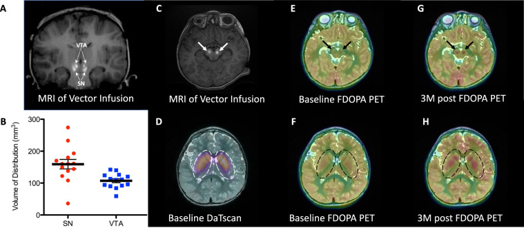
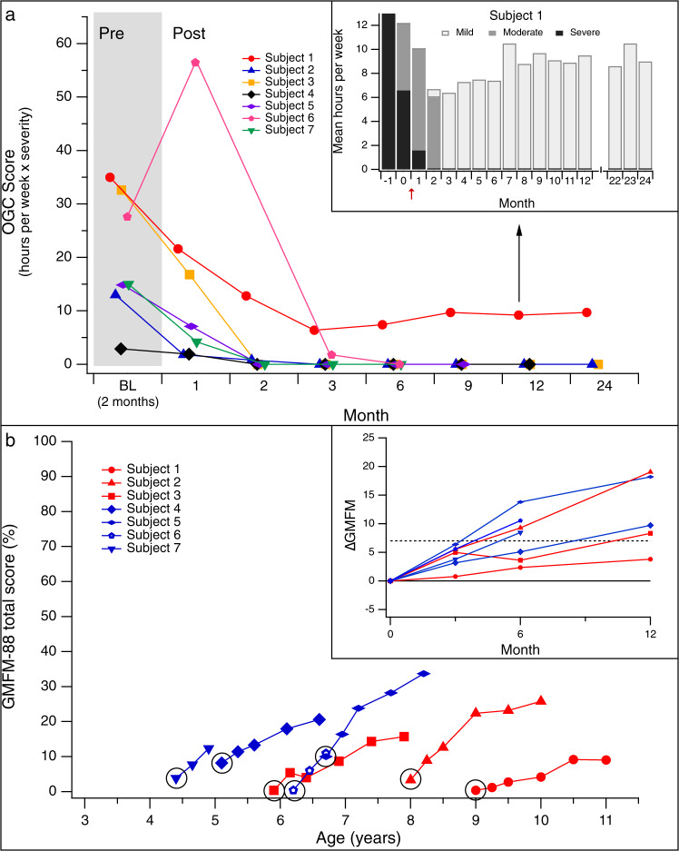
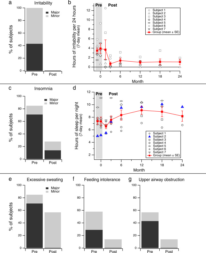
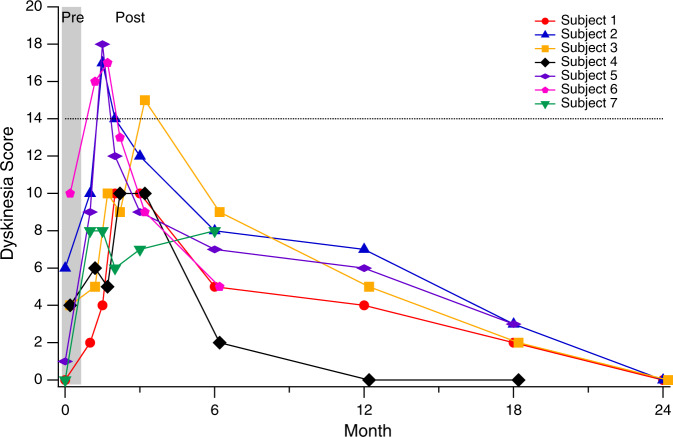
Aromatic L-amino acid decarboxylase (AADC) deficiency is a rare genetic disorder characterized by deficient synthesis of dopamine and serotonin. It presents in early infancy, and causes severe developmental disability and lifelong motor, behavioral, and autonomic symptoms including oculogyric crises (OGC), sleep disorder, and mood disturbance. We investigated the safety and efficacy of delivery of a viral vector expressing AADC (AAV2-hAADC) to the midbrain in children with AADC deficiency (ClinicalTrials.gov Identifier NCT02852213). Seven (7) children, aged 4-9 years underwent convection-enhanced delivery (CED) of AAV2-hAADC to the bilateral substantia nigra (SN) and ventral tegmental area (VTA) (total infusion volume: 80 µL per hemisphere) in 2 dose cohorts: 1.3 × 10¹¹ vg (n = 3), and 4.2 × 10¹¹ vg (n = 4). Primary aims were to demonstrate the safety of the procedure and document biomarker evidence of restoration of brain AADC activity. Secondary aims were to assess clinical improvement in symptoms and motor function. Direct bilateral infusion of AAV2-hAADC was safe, well-tolerated and achieved target coverage of 98% and 70% of the SN and VTA, respectively. Dopamine metabolism was increased in all subjects and FDOPA uptake was enhanced within the midbrain and the striatum. OGC resolved completely in 6 of 7 subjects by Month 3 post-surgery. Twelve (12) months after surgery, 6/7 subjects gained normal head control and 4/7 could sit independently. At 18 months, 2 subjects could walk with 2-hand support. Both the primary and secondary endpoints of the study were met. Midbrain gene delivery in children with AADC deficiency is feasible and safe, and leads to clinical improvements in symptoms and motor function.


Conflict of interest statement

N.G. reports relationships with Oscine Therapeutics (consulting) and Y-mAbs Therapeutics (consulting). K.H. reports that he is employed by Medical Neurogenetics Laboratories, a company that provides commercial diagnostic testing for aromatic l-amino acid decarboxylase deficiency. P.L. reports relationships with Axovant (Advisory Board), Neurocrine Biosciences (research funding, consulting), UniQure (research funding), Voyager Therapeutics (research funding), and Clearpoint Neuro (consulting). K.S.B. is the founder and equity holder of Brain Neurotherapy Bio. The remaining authors have no competing interests to disclose.

Figures

Fig. 1
Fig. 1. MR-guided delivery of AAV2-hAADC into the midbrain, baseline DaTscan and changes in FDOPA PET biomarker after gene delivery.
Coronal (A) and axial (C) MR images at the conclusion of the vector infusions into SN and VTA regions (white arrows). Bright signal corresponds to gadoteridol admixed with AAV2-hAADC. Infusions are performed sequentially while imaging, starting with right SN. Please, note accurate targeting and coverage in respective anatomical regions. B Coverage (volume of distribution, Vd) of all infusions performed into the SN and VTA in 7 subjects (n = 2 independent infusion sites (left and right) examined per participant (n = 7) for each target structure (SNc and VTA), for a total of n = 14 independent infusions per target structure). SN infusion (50 µL) achieved coverage of ~160 ± 60 mm3 (mean ± SD, n = 14 infusions (two infusions per participant)) with one suboptimal infusion due to leakage along the perivascular space. VTA infusion (30 µL) resulted in coverage of 103 ± 22 mm3. Coverage volume of gadoteridol (Vd) suggests almost 80% anatomical coverage of both SN and VTA in all subjects (except single SN in one patient). D DaTscan imaging of the striatum at baseline confirmed a normal pattern of dopaminergic innervation in all study subjects, indicative of preserved nigrostriatal pathway. E, F Baseline FDOPA imaging of the midbrain regions (SN and VTA, black arrows in E) and nigrostriatal projection (caudate nucleus and putamen, dotted line in F). Lack of signal in both regions represents impaired conversion of FDOPA to F-dopamine due to absent AADC activity. G, H Increased FDOPA PET uptake 3 months after AAV2-hAADC administration in the midbrain and striatum, respectively. Images for Subject 4 are shown as representative of the group; see Supplementary Fig. S1 for images for each individual subject. Source data are provided as a Source Data file.
Fig. 2
Fig. 2. CSF Neurotransmitter metabolites.
Concentrations of CSF metabolites measured at 2 separate baseline (BL) timepoints, Month 3, Month 6, and Month 12, in individual subjects in Cohort 1 (low-dose, black markers); and Cohort 2 (high-dose, white markers) and summarized at each timepoint as the group median (bars). A Homovanillic acid (HVA), the dopamine metabolite, was significantly higher at each post-operative timepoint compared to the baseline mean (inverted bracket; (*p = 0.0078 at Months 3 and 6, p = 0.0313 at Month 12, one-tailed Wilcoxon signed-rank test). Lower limit of normal range: 218 nmol/L (dotted line). B 5-hydroxyindoleacetic acid (5-HIAA), the serotonin metabolite, did not change after gene delivery. Lower limit of normal range: 66 nmol/L (dotted line). C 3-O-methyldopa (3-OMD) was elevated in all subjects at all timepoints. Normal: <100 nmol/L (dotted line). Source data can be found in Table 2.
Fig. 3
Fig. 3. Changes in Oculogyric Crises (OGC) and motor function after gene delivery.
A The OGC score was calculated monthly, and represents the weekly average of the duration of episodes (hours per week), weighted by severity (grade 1–3: 1 = mild/eye deviation only; 2 = moderate/eye deviation + dystonia or dyskinesia of the face and/or neck; 3 = severe/dystonia or dyskinesia involving the trunk and/or limbs). By Month 3, OGCs completely resolved in 6/7 subjects. Inset (black arrow): Subject 1 had residual episodes throughout the 24-month study period; the severity decreased after surgery (red arrow). B Gross Motor Function Measure-88 (GMFM-88) scores for Cohorts 1 (red) and 2 (blue). Data are shown through Month 24 for Subjects 1–3, Month 18 for Subjects 4 and 5, and Month 6 for Subjects 6 and 7. Baseline GMFM scores for all subjects (circled) were ≤10, consistent with severe motor impairment. Inset: changes in GMFM score between Baseline and Month 12. An increase of ≥7 points (dotted line, representing a clinically meaningful positive change) was observed in 6/7 subjects by Month 12. Source data are provided as a Source Data file.
Fig. 4
Fig. 4. Non-motor symptoms before and after gene delivery.
A The percentage of subjects (n = 7) who, by caregiver report, experienced irritability at baseline compared to the last available follow-up timepoint (Month 24 for Subjects 1–3, Month 18 for Subjects 4 and 5, and Month 6 for Subjects 6 and 7). B The number of hours or irritability per day recorded in caregiver diaries increased slightly immediately after surgery, and then improved on average (mean ± SE). C Prevalence and severity of insomnia before and after gene delivery by caregiver report. D The mean number of hours (±SE) of night-time sleep recorded in caregiver diaries improved slightly across subjects after gene delivery. For Subject 2 (blue triangles), the improvement was dramatic. EG Prevalence and severity of other non-motor symptoms before and after gene delivery by caregiver report (see details in A, above). Major (black): frequent and/or severe, with significant impact on comfort or function; Minor (gray): infrequent and/or mild. Source data are provided as a Source Data file.
Fig. 5
Fig. 5. Time course of dyskinesia after gene transfer.
Dyskinesia score, adapted from the Abnormal Involuntary Movement Scale (AIMS). Involuntary movements in each of 7 body regions were scored from 0 (none) to 4 (severe), for a maximum possible score of 28. A total score of 14 (dotted line) may correspond with either mild generalized dyskinesia, or moderate-severe involuntary movements in a more focal distribution. Dyskinesia peaked in severity between 1 and 3 months after surgery, and gradually improved thereafter. At Month 24, dyskinesia had resolved completely in all 3 subjects in Cohort 1.

References

    1. Hyland K, Clayton PT. Aromatic amino acid decarboxylase deficiency in twins. J. Inherit. Metab. Dis. 1990;13:301–304. doi: 10.1007/BF01799380. - DOI - PubMed
    1. Wassenberg T, et al. Consensus guideline for the diagnosis and treatment of aromatic l-amino acid decarboxylase (AADC) deficiency. Orphanet J. Rare Dis. 2017;12:12. doi: 10.1186/s13023-016-0522-z. - DOI - PMC - PubMed
    1. Brun L, et al. Clinical and biochemical features of aromatic L-amino acid decarboxylase deficiency. Neurology. 2010;75:64–71. doi: 10.1212/WNL.0b013e3181e620ae. - DOI - PubMed
    1. Pearson, T. S. et al. AADC deficiency from infancy to adulthood: symptoms and developmental outcome in an international cohort of 63 patients. J. Inherit. Metab. Dis. (2020) 10.1002/jimd.12247. - PMC - PubMed
    1. Hwu, W. L., Chien, Y. H., Lee, N. C. & Li, M. H. Natural history of aromatic L-amino acid decarboxylase deficiency in Taiwan. JIMD Rep. (2017) 10.1007/8904_2017_54. - PMC - PubMed

Publication types

MeSH terms

Substances

Supplementary concepts

Associated data