

Changes in the avascular area of the meniscus using mesenchymal stem cells and growth plate chondrocytes in a pig model
- PMID: 34254669
- PMCID: PMC8602013
- DOI: 10.1111/joa.13508
Changes in the avascular area of the meniscus using mesenchymal stem cells and growth plate chondrocytes in a pig model
Abstract
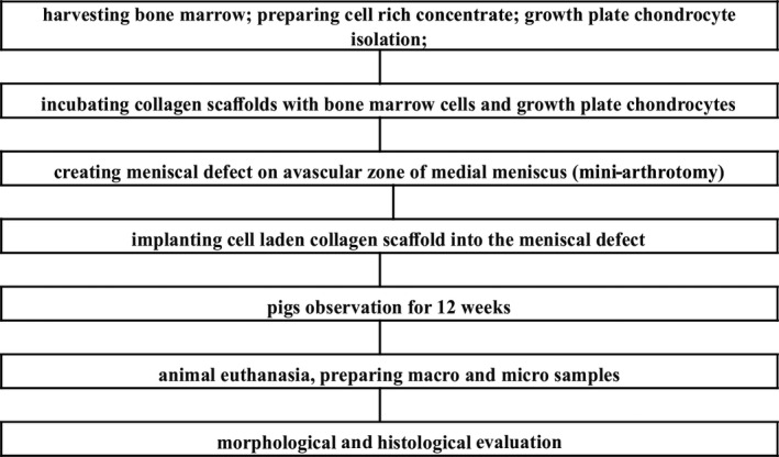
Menisci are wedge-shaped cartilage discs that are divided into two parts: the avascular and vascular regions. They are formed by fibrocartilage tissue, which contains round cartilage-like cells and extracellular matrix. Meniscus injury in animals is a common orthopedic problem, but data on the natural healing process mainly deals with the vascular zone. The healing processes in the avascular zone of the meniscus are significantly limited. Thus, this study aimed to evaluate autologous growth plate chondrocytes' impact on the healing process of a damaged meniscus in the avascular zone based on a growing animal model. The study group consisted of 10 pigs at about three months of age. From each animal, chondrocytes from the iliac growth plate and from concentrated bone marrow were taken. Knee joints were divided into right (R) and left (L). The medial meniscus of the R knee joint was treated with a hyaluronic acid based scaffold incubated with bone marrow cells from marrow aspirates (nCHON). The medial meniscus of the L knee joint was treated with a hyaluronic acid based scaffold incubated with bone marrow cells from marrow aspirates supplemented with immature chondrocytes isolated from growth plates (wCHON). The meniscus was damaged in the avascular zone in both knee joints. Followingly, the damaged part of the meniscus was filled with a scaffold with cells from the concentrated bone marrow and from growth plate chondrocytes. In the control group, a scaffold with concentrated bone marrow cells was used. After three months the animals were euthanized and preparations (microscopic slides) were made from the meniscus' damaged part. A qualitative and quantitative analysis have been prepared. The wCHON group in comparison with the nCHON group showed a statistically significantly higher number of fusiform cells on the surface of the graft as well as better healing of the graft. In addition, the degree of vascularization was higher in specimens from the wCHON group than in the nCHON group. The results of our research on immature pig knees revealed that mesenchymal stem cell and growth plate chondrocytes could be treated as the cell source for meniscus reconstruction, and growth plate chondrocytes enhance healing processes in the avascular zone of the injured meniscus.
Keywords: growth plate chondrocytes; meniscus tear; mesenchymal stem cells; pigs; scaffold.
© 2021 Anatomical Society.
Conflict of interest statement
Author declare that they do not have any conflict of interests.
Figures


Similar articles
-
Enhancement of cartilage repair through the addition of growth plate chondrocytes in an immature skeleton animal model.J Orthop Surg Res. 2019 Aug 15;14(1):260. doi: 10.1186/s13018-019-1302-y. J Orthop Surg Res. 2019. PMID: 31416470 Free PMC article.
-
Stem cell-based tissue-engineering for treatment of meniscal tears in the avascular zone.J Biomed Mater Res B Appl Biomater. 2013 Oct;101(7):1133-42. doi: 10.1002/jbm.b.32922. Epub 2013 Apr 6. J Biomed Mater Res B Appl Biomater. 2013. PMID: 23564690
-
[Treatment of a bone bridge by transplantation of mesenchymal stem cells and chondrocytes in a composite scaffold in pigs: experimental study].Acta Chir Orthop Traumatol Cech. 2011;78(6):528-36. Acta Chir Orthop Traumatol Cech. 2011. PMID: 22217406 Czech.
-
Locally applied angiogenic factors--a new therapeutic tool for meniscal repair.Ann Anat. 2005 Nov;187(5-6):509-19. doi: 10.1016/j.aanat.2005.04.010. Ann Anat. 2005. PMID: 16320830 Review.
-
Tissue engineering of the meniscus.Biomaterials. 2004 Apr;25(9):1523-32. doi: 10.1016/s0142-9612(03)00499-x. Biomaterials. 2004. PMID: 14697855 Review.
Cited by
-
Advancements in the treatment of osteochondral lesions of the talus.J Orthop Surg Res. 2024 Dec 6;19(1):827. doi: 10.1186/s13018-024-05314-6. J Orthop Surg Res. 2024. PMID: 39639331 Free PMC article. Review.
References
-
- Al Faqeh, H. , Nor Hamdan, B.M. , Chen, H.C. , Aminuddin, B.S. & Ruszymah, B.H. (2012) The potential of intra‐articular injection of chondrogenic‐induced bone marrow stem cells to retard the progression of osteoarthritis in a sheep model. Experimental Gerontology, 47, 658–664. - PubMed
-
- Amann, E. , Wolff, P. , Breel, E. , van Griensven, M. & Balmayor, E.R. (2017) Hyaluronic acid facilitates chondrogenesis and matrix deposition of human adipose derived mesenchymal stem cells and human chondrocytes co‐cultures. Acta Biomaterialia, 52, 130–144. - PubMed
-
- Angele, P. , Johnstone, B. , Kujat, R. , Zellner, J. , Nerlich, M. , Goldberg, V. et al. (2008) Stem cell based tissue engineering for meniscus repair. Journal of Biomedical Materials Research Part A, 85, 445–455. - PubMed
-
- Beaufils, P. & Pujol, N. (2018) Meniscal repair: technique. Orthopaedics and Traumatology: Surgery and Research, 104, 137–145. - PubMed
Publication types
MeSH terms
LinkOut - more resources
Full Text Sources
