. 2021 Aug 31;6(4):e0029321.
doi: 10.1128/mSystems.00293-21. Epub 2021 Jul 13.

High Transcriptional Activity and Diverse Functional Repertoires of Hundreds of Giant Viruses in a Coastal Marine System

Affiliations

High Transcriptional Activity and Diverse Functional Repertoires of Hundreds of Giant Viruses in a Coastal Marine System

Anh D Ha et al. mSystems. .

Abstract

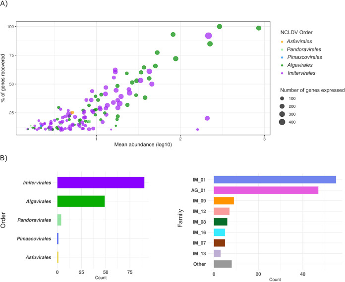
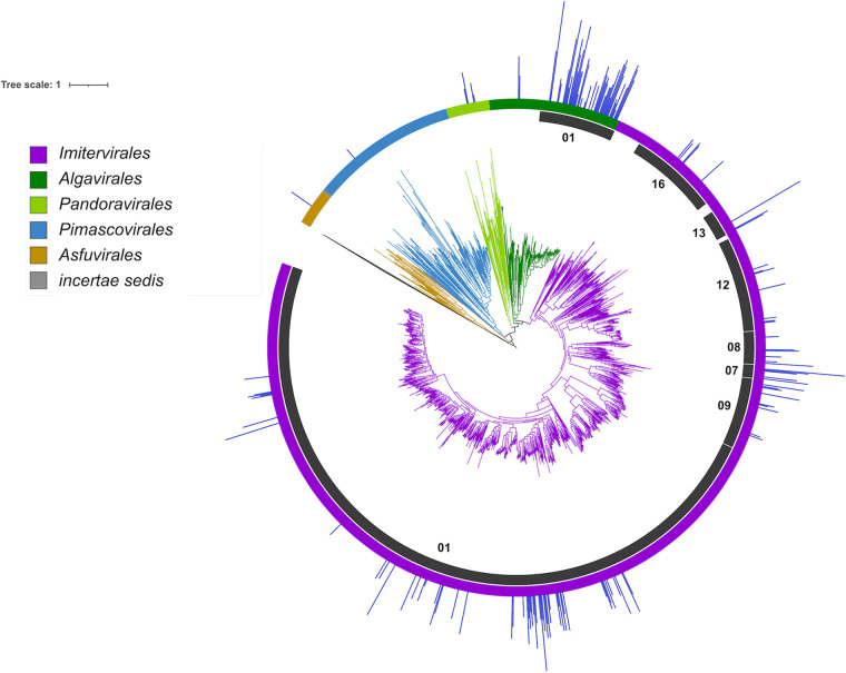
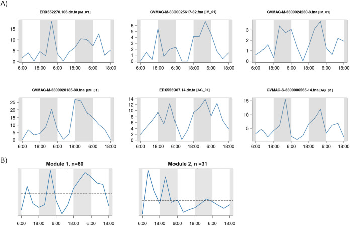
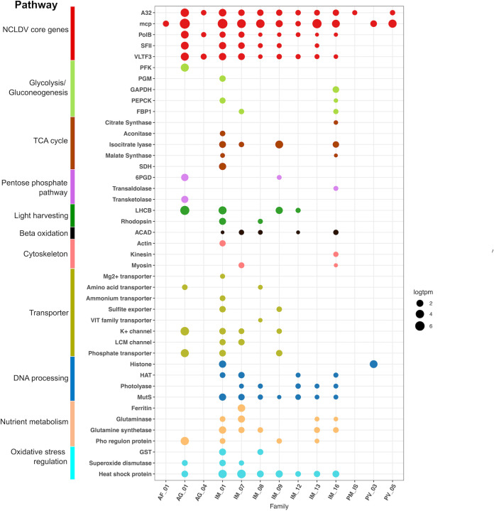
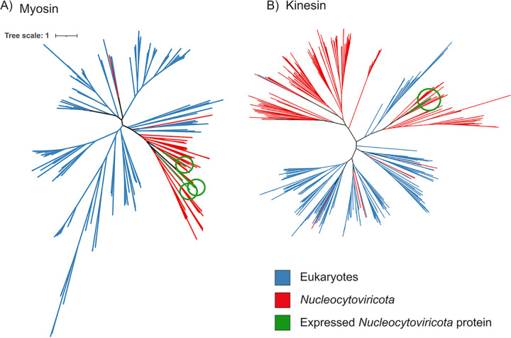
Viruses belonging to the Nucleocytoviricota phylum are globally distributed and include members with notably large genomes and complex functional repertoires. Recent studies have shown that these viruses are particularly diverse and abundant in marine systems, but the magnitude of actively replicating Nucleocytoviricota present in ocean habitats remains unclear. In this study, we compiled a curated database of 2,431 Nucleocytoviricota genomes and used it to examine the gene expression of these viruses in a 2.5-day metatranscriptomic time-series from surface waters of the California Current. We identified 145 viral genomes with high levels of gene expression, including 90 Imitervirales and 49 Algavirales viruses. In addition to recovering high expression of core genes involved in information processing that are commonly expressed during viral infection, we also identified transcripts of diverse viral metabolic genes from pathways such as glycolysis, the TCA cycle, and the pentose phosphate pathway, suggesting that virus-mediated reprogramming of central carbon metabolism is common in oceanic surface waters. Surprisingly, we also identified viral transcripts with homology to actin, myosin, and kinesin domains, suggesting that viruses may use these gene products to manipulate host cytoskeletal dynamics during infection. We performed phylogenetic analysis on the virus-encoded myosin and kinesin proteins, which demonstrated that most belong to deep-branching viral clades, but that others appear to have been acquired from eukaryotes more recently. Our results highlight a remarkable diversity of active Nucleocytoviricota in a coastal marine system and underscore the complex functional repertoires expressed by these viruses during infection. IMPORTANCE The discovery of giant viruses has transformed our understanding of viral complexity. Although viruses have traditionally been viewed as filterable infectious agents that lack metabolism, giant viruses can reach sizes rivalling cellular lineages and possess genomes encoding central metabolic processes. Recent studies have shown that giant viruses are widespread in aquatic systems, but the activity of these viruses and the extent to which they reprogram host physiology in situ remains unclear. Here, we show that numerous giant viruses consistently express central metabolic enzymes in a coastal marine system, including components of glycolysis, the TCA cycle, and other pathways involved in nutrient homeostasis. Moreover, we found expression of several viral-encoded actin, myosin, and kinesin genes, indicating viral manipulation of the host cytoskeleton during infection. Our study reveals a high activity of giant viruses in a coastal marine system and indicates they are a diverse and underappreciated component of microbial diversity in the ocean.

Keywords: NCLDV; Nucleocytoviricota; cytoskeleton; giant viruses; kinesin; myosin; viral diversity.


Figures

FIG 1
FIG 1
(A) General statistics of the 145 Nucleocytoviricota genomes. Each dot represents a viral genome; the x axis shows the mean abundance of the genome across 16 time points (TPM, log10 transformed); the y axis shows the percentage of genes in a viral genome that were recovered across all samples; and the dot size is scaled to the total number of genes recovered. (B) The taxonomic distribution of the 145 genomes. The x axis represents the total number of viruses in each taxon, and the y axis shows the Nucleocytoviricota order (left) and family (right). In the family plot (right), families with less than three members are collapsed down into the “Other” category. Abbreviations: IM, Imitervirales; AG, Algavirales.
FIG 2
FIG 2
Phylogeny of Nucleocytoviricota with mean read mapping (TPM, natural log transformed) on the outermost bar plot. The order-level classification of Nucleocytoviricota is denoted by the branch colors and the outer color strip, and particular families of interest are highlighted in the inner ring. IM_01 corresponds to the Mesomimiviridae, IM_16 corresponds to the Mimiviridae, and AG_01 corresponds to the Prasinoviridae.
FIG 3
FIG 3
Expression levels of Nucleocytoviricota during the 16 time points studied. (A) Whole-genome expression profiles of six genomes with significant difference in expression level between daytime and nighttime (Mann-Whitney U test P < 0.1). The x axis represents the 16 time points and the y axis represents the total expression (TPM). Nucleocytoviricota family classifications are shown in brackets. Abbreviations: IM, Imitervirales; AG, Algavirales. (B) Eigengene of the genome coexpression modules. The x axis represents the 16 time points and the y axis represents the eigengene expression values (arbitrary units).
FIG 4
FIG 4
Metabolic genes expressed in the transcriptomes. The x axis shows different viral clades and the y axis denotes the functional annotation. The sizes of the bubbles represent the total abundance of the gene in TPM (natural log transformed) and the colors show the functional category. Family assignment abbreviations: AF, Asfuvirales; IM, Imitervirales; AG, Algavirales; PM, Pimascovirales; PV, Pandoravirales; IS, incertae sedis. Gene function abbreviations: PFK, phosphofructokinase; PGM, phosphoglycerate mutase; GAPDH, glyceraldehyde 3-P dehydrogenase; PEPCK, phosphoenolpyruvate carboxykinase; FBP1, fructose-1-6-bisphosphatase; SDH, succinate dehydrogenase; 6PGD, 6-phosphogluconate dehydrogenase; LHCB, chlorophyll a/b binding protein; ACAD, acyl-CoA dehydrogenase; HAT, histone acetyltransferase; GST, glutathione S-transferase.
FIG 5
FIG 5
Phylogeny of Nucleocytoviricota myosin (left) and kinesin (right) proteins together with references available in the EggNOG database. The myosin and kinesin genes that were identified in the metatranscriptomes are colored green.
FIG 6
FIG 6
Host-virus expression correlation network. Each node represents a eukaryotic genome (circle) or viral genome (square). The lines between two genomes denote a correlation coefficient larger than 0.8. The sizes of the nodes represent the total abundance in TPM (natural log transformed) of the genome and the colors show the genome’s taxonomy.

References

    1. Fischer MG. 2016. Giant viruses come of age. Curr Opin Microbiol 31:50–57. doi:10.1016/j.mib.2016.03.001. - DOI - PubMed
    1. Koonin EV. 2005. Virology: Gulliver among the Lilliputians. Curr Biol 15:R167–R169. doi:10.1016/j.cub.2005.02.042. - DOI - PubMed
    1. Koonin EV, Yutin N. 2010. Origin and evolution of eukaryotic large nucleo-cytoplasmic DNA viruses. Intervirology 53:284–292. doi:10.1159/000312913. - DOI - PMC - PubMed
    1. Koonin EV, Dolja VV, Krupovic M, Varsani A, Wolf YI, Yutin N, Zerbini FM, Kuhn JH. 2020. Global organization and proposed megataxonomy of the virus world. Microbiol Mol Biol Rev 84:e00061-19. doi:10.1128/MMBR.00061-19. - DOI - PMC - PubMed
    1. Aylward FO, Moniruzzaman M, Ha AD, Koonin EV. 2021. A phylogenomic framework for charting the diversity and evolution of giant viruses. bioRxiv doi:10.1101/2021.05.05.442809. - DOI - PMC - PubMed

LinkOut - more resources