

Assessment of tumor depth in oral tongue squamous cell carcinoma with multiparametric MRI: correlation with pathology
- PMID: 34255162
- PMCID: PMC8660735
- DOI: 10.1007/s00330-021-08148-6
Assessment of tumor depth in oral tongue squamous cell carcinoma with multiparametric MRI: correlation with pathology
Abstract
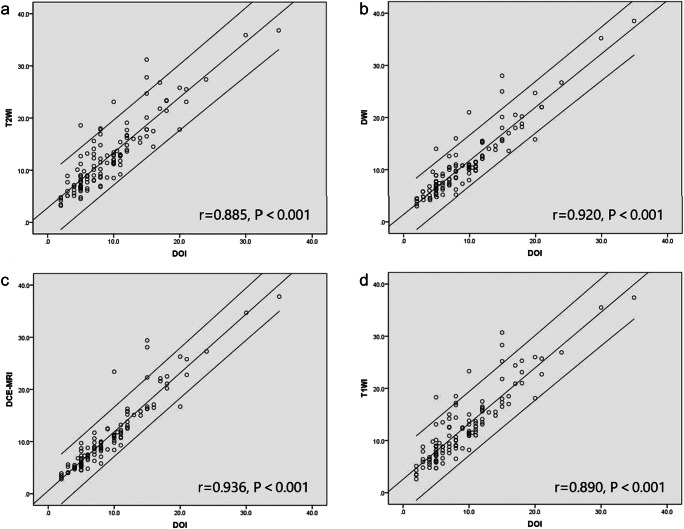
Objectives: To compare the correlation of depth of invasion (DOI) measured on multiple magnetic resonance imaging (MRI) sequences and pathological DOI, in order to determine the optimal MRI sequence for measurement.
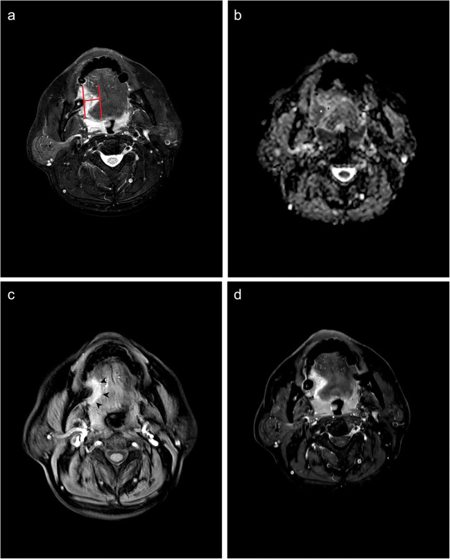
Methods: A total of 122 oral tongue squamous cell carcinoma (OTSCC) patients were retrospectively analyzed, who had received preoperative MRI and surgical resection. DOIs measured on fat-suppressed T2-weighted imaging (T2WI), diffusion-weighted imaging (DWI), dynamic enhanced-T1 high-resolution insotropic volume examination (e-THRIVE), and contrast-enhanced fat-suppressed T1WI (CE-T1WI) were respectively compared to those measured in pathologic specimens. The cutoff value of the best correlated MRI sequence was determined, and the T staging accuracy of MRI-derived DOI was evaluated.
Results: DOI derived from e-THRIVE showed the best correlation (r = 0.936, p < 0.001) with pathological DOI. The area under the curve values of MRI-derived DOI distinguishing T1 stage from T2 stage and distinguishing T2 stage from T3 stage were 0.969 and 0.974, respectively. The T staging criteria of MRI-derived DOI were 6.2 mm and 11.4 mm, with a staging accuracy of 86.9% compared to pathological DOI criteria of 5 mm and 10 mm.
Conclusion: E-THRIVE was the optimal MR sequence to measure the MR-derived DOI, and DOI derived from e-THRIVE could serve as a potential cut-off value as a clinical T staging indicator of OTSCC.
Key points: • Multiparametric MRI helps radiologists to assess the neoplasm invasion in patients with oral tongue squamous cell carcinoma. • Retrospective study indicated that measurement was most accurate on enhanced-T1 high-resolution insotropic volume examination dynamic contrast enhancement images. • T staging of oral tongue squamous cell carcinoma was accurate according to the dynamic contrast enhancement MRI-derived depth of invasion.
Keywords: Multiparametric MRI; Neoplasm Invasion; Oral tongue squamous cell carcinoma.
© 2021. The Author(s).
Conflict of interest statement
The authors of this manuscript declare no relationships with any companies whose products or services may be related to the subject matter of the article.
Figures

Similar articles
-
Utility of Diffusion-weighted MR Imaging for Evaluating the Depth of Invasion in Oral Tongue Squamous Cell Carcinoma.Magn Reson Med Sci. 2025 Apr 1;24(2):210-219. doi: 10.2463/mrms.mp.2023-0137. Epub 2024 Mar 7. Magn Reson Med Sci. 2025. PMID: 38447989 Free PMC article.
-
Value of radiological depth of invasion in non-pT4 Oral tongue squamous cell carcinoma: implication for preoperative MR T-staging.Eur Radiol. 2024 Sep;34(9):6047-6059. doi: 10.1007/s00330-024-10598-7. Epub 2024 Feb 3. Eur Radiol. 2024. PMID: 38308013 Free PMC article.
-
Usefulness of contrast-enhanced CT in the evaluation of depth of invasion in oral tongue squamous cell carcinoma: comparison with MRI.Oral Radiol. 2021 Jan;37(1):86-94. doi: 10.1007/s11282-020-00429-y. Epub 2020 Feb 21. Oral Radiol. 2021. PMID: 32086730
-
The accuracy of magnetic resonance imaging to measure the depth of invasion in oral tongue cancer: a systematic review and meta-analysis.Int J Oral Maxillofac Surg. 2022 Apr;51(4):431-440. doi: 10.1016/j.ijom.2021.07.010. Epub 2021 Aug 20. Int J Oral Maxillofac Surg. 2022. PMID: 34420832
-
The prognostic role of the pre-treatment neutrophil to lymphocyte ratio (NLR) and tumor depth of invasion (DOI) in early-stage squamous cell carcinomas of the oral tongue.Oral Maxillofac Surg. 2022 Mar;26(1):21-32. doi: 10.1007/s10006-021-00969-5. Epub 2021 Jun 9. Oral Maxillofac Surg. 2022. PMID: 34106358 Review.
Cited by
-
Foreign body granuloma in the tongue differentiated from tongue cancer: A case report.World J Clin Cases. 2022 Jun 26;10(18):6247-6253. doi: 10.12998/wjcc.v10.i18.6247. World J Clin Cases. 2022. PMID: 35949813 Free PMC article.
-
MicroRNA-Based Markers of Oral Tongue Squamous Cell Carcinoma and Buccal Squamous Cell Carcinoma: A Systems Biology Approach.Biochem Res Int. 2023 Apr 24;2023:5512894. doi: 10.1155/2023/5512894. eCollection 2023. Biochem Res Int. 2023. PMID: 37143570 Free PMC article.
-
Prognostic value of radiological T category using conventional MRI in patients with oral tongue cancer: comparison with pathological T category.Neuroradiology. 2024 Jun;66(6):907-917. doi: 10.1007/s00234-024-03345-8. Epub 2024 Apr 12. Neuroradiology. 2024. PMID: 38607437 Free PMC article.
-
Accuracy of Magnetic Resonance Imaging in Detecting Tumor Depth of Invasion in Squamous Cell Carcinoma of the Tongue: A Systematic Review.J Maxillofac Oral Surg. 2023 Sep;22(3):720-727. doi: 10.1007/s12663-023-01886-8. Epub 2023 Apr 25. J Maxillofac Oral Surg. 2023. PMID: 37534361 Free PMC article. Review.
-
Utility of Diffusion-weighted MR Imaging for Evaluating the Depth of Invasion in Oral Tongue Squamous Cell Carcinoma.Magn Reson Med Sci. 2025 Apr 1;24(2):210-219. doi: 10.2463/mrms.mp.2023-0137. Epub 2024 Mar 7. Magn Reson Med Sci. 2025. PMID: 38447989 Free PMC article.
References
MeSH terms
Grants and funding
LinkOut - more resources
Full Text Sources
Medical
