Review
. 2021 Jul 13:23:1-27.
doi: 10.1146/annurev-bioeng-092419-060810.

Vascular Mechanobiology: Homeostasis, Adaptation, and Disease

Affiliations
Review

Vascular Mechanobiology: Homeostasis, Adaptation, and Disease

Jay D Humphrey et al. Annu Rev Biomed Eng. .

Abstract

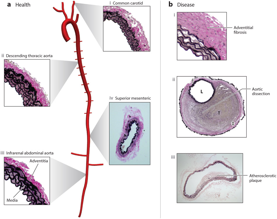
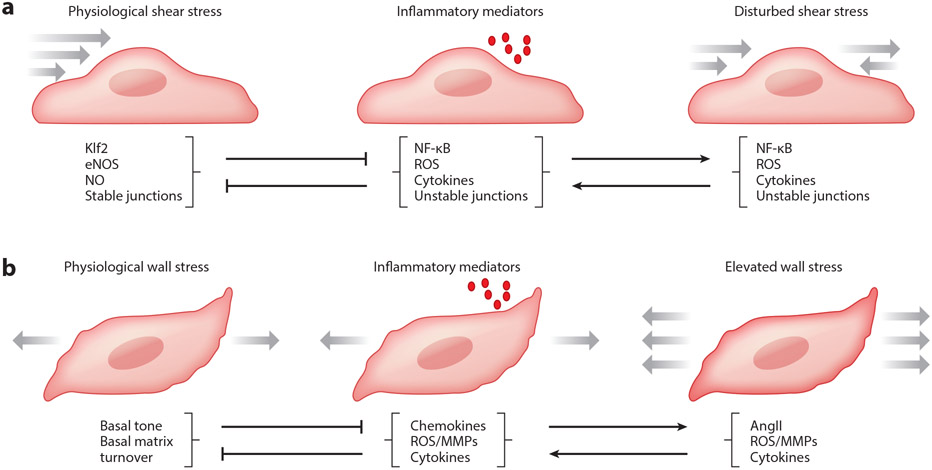
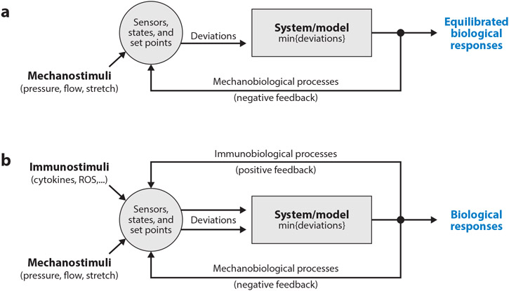
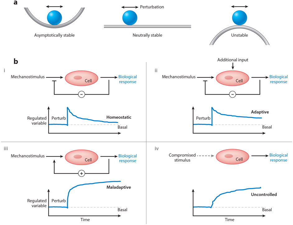
Cells of the vascular wall are exquisitely sensitive to changes in their mechanical environment. In healthy vessels, mechanical forces regulate signaling and gene expression to direct the remodeling needed for the vessel wall to maintain optimal function. Major diseases of arteries involve maladaptive remodeling with compromised or lost homeostatic mechanisms. Whereas homeostasis invokes negative feedback loops at multiple scales to mediate mechanobiological stability, disease progression often occurs via positive feedback that generates mechanobiological instabilities. In this review, we focus on the cell biology, wall mechanics, and regulatory pathways associated with arterial health and how changes in these processes lead to disease. We discuss how positive feedback loops arise via biomechanical and biochemical means. We conclude that inflammation plays a central role in overriding homeostatic pathways and suggest future directions for addressing therapeutic needs.

Keywords: aneurysms; atherosclerosis; feedback; fibrosis; hypertension; mechanotransduction.


Figures

Figure 1
Figure 1
(a) Overview of central arteries of the mouse vasculature reconstructed from a microCT image, showing the aorta, brachiocephalic, common carotid, subclavian, intercostal, and renal arteries, with representative histological images of portions of (i) a healthy common carotid artery, (ii) descending thoracic aorta, and (iii) infrarenal abdominal aorta (all elastic) as well as (iv) superior mesenteric artery (muscular). Note the nearly concentric elastic lamellar structures in the media of the elastic arteries (elastin shown black) in contrast to the smooth-muscle rich media of the muscular artery. Note, too, the abundant collagen (pink) in the adventitia of both types of arteries. Arterial microstructure arises during development and is maintained in health via a continual replacement of cells that die and matrix that degrades, accomplished via homeostatic processes driven by negative feedback. Resident macrophages can participate in the homeostatic removal of dead cells, debris, and damaged matrix. (b) In contrast, cross-sections are shown for three different pathologies that affect the large arteries: (i) hypertension induced adventitial fibrosis, (ii) contained rupture with a thrombus (T) filled false lumen, with L denoting the true lumen and daggers the adventitia, and (iii) an early atherosclerotic lesion in the intima of the abdominal aorta. Such maladaptive remodeling or catastrophic events are driven and/or preceded by compromised homeostasis, often via positive feedback.
Figure 2
Figure 2
(a) Schema of endothelial cells subjected to mechanical and/or inflammatory stimuli. Physiological levels of wall shear stress induced by blood flow activate multiple pathways that oppose inflammation and remodeling, most prominently Klf2, which mediates expression of anti-inflammatory, anti-thrombotic, and anti-oxidative genes. One important downstream gene is eNOS, which catalyzes the synthesis of NO, which promotes junctional stability, suppresses SMC contraction and proliferation, and suppresses activation of leukocytes and platelets. By contrast, disturbed flow or inflammatory mediators activate the transcription factor NF-kB and downstream genes, inducing reactive oxygen species (ROS) production and destabilizing junctions. NF-kB reduces Klf2 expression, and ROS react with NO to generate peroxynitrite. High levels of inflammatory mediators over long periods suppress expression of flow sensors such as PECAM and VE-cadherin, which inhibit flow responses. Disturbed flow also sensitizes ECs to inflammatory factors, amplifying these responses. (b) Schema of smooth muscle cells subjected to mechanical and/or inflammatory stimuli. Physiological levels of intramural stresses arising largely from blood pressure stimulate continual turnover of cells and matrix to maintain the structure and function of the arterial wall despite inevitable cell death and matrix degradation over long periods in maturity. Both elevated wall stress, as in hypertension, and inflammatory cell infiltration can disrupt normal processes and lead to maladaptive remodeling or diverse disease states (cf. Figure 1). The latter are driven via the increased production or presence of chemokines (such as MCP-1), cytokines (such as TGFβ, IL-6, IL-17a, and TNF-α), and matrix metalloproteinases (such as MMP-1, 2, 9, 12).
Figure 3
Figure 3
(a) Schema of key components of mechanical homeostasis in arteries. Mechanical stimuli (pressure, flow, and axial stretch) combine with local geometry and properties to define the mechanical state, which can be calculated using equations of continuum biomechanics. Cells must sense this state and compare perceived values against homeostatic targets, or set-points. Any deviation in mechanical state from the set-point is then used by the effector cells to modify their phenotype and/or the extracellular matrix (collectively the system to be modeled). Various models, both phenomenological and mechanistic, single-scale and multi-scale, can be used to determine the associated biological response, which can affect the geometry and material properties and hence the mechanical state. Continued negative feedback can maintain or restore the homeostatic state. (b) Although (para-) inflammation can contribute to the negative feedback that is dominated in maturity by the mechanobiology, chronic inflammation often drives a positive feedback that exacerbates the perturbation and drives disease progression. Various cell types can contribute to this disease-promoting inflammation, including monocytes/macrophages and T-cells though also ECs, SMCs, or FBs that undergo a phenotypic modulation towards an inflammatory (or degradative) phenotype.
Figure 4
Figure 4
(a) Conceptual representation of three common types of (in)stabilities: asymptotically stable, neutrally stable, and unstable. Imagine, respectively, that a perturbation in loading would cause the rigid sphere to move, returning to its original location on the left, finding a new nearby location in the middle, and falling far from its original position on the right. Note that neutral stability allows adaptivity in biology, but also increased vulnerability to instabilities that manifest as diseases. (b) Possible control systems relevant to arterial health and disease: three show responses to perturbations (vertical change in the regulated variable, such as stress or material stiffness) and one (lower right) in the context of compromised mechano-sensing. In particular, negative feedback (−) is restorative, as in cases of acute or modest sustained changes in hemodynamics (upper left), but can be over-ridden by additional stimuli, such as inflammation (upper right). In contrast, positive feed-back (+), as in adventitial fibrosis (cf. Figure 1), is typically maladaptive, moving the controlled variable from its basal value (horizontal dashed line). Finally, systems that rely on appropriate input can become maladaptive when the input signal is compromised or lost, as in dysfunctional mechanotransduction. Overall, negative feedback promotes asymptotic mechanobiological stability, negative feedback with additional non-mechanical stimuli represents neutral stability, and cases of positive feedback or compromised mechano-input represent instabilities.

References

    1. Dajnowiec D, Langille BL. 2007. Arterial adaptations to chronic changes in haemodynamic function: coupling vasomotor tone to structural remodelling. Clin Sci (Lond) 113:15–23 - PubMed
    1. Bikfalvi A 2006. Angiogenesis: health and disease. Ann Oncol 17 Suppl 10:x65–70 - PubMed
    1. Humphrey JD, Dufresne ER, Schwartz MA. 2014. Mechanotransduction and extracellular matrix homeostasis. Nat Rev Mol Cell Biol 15:802–12 - PMC - PubMed
    1. Safar ME. 2010. Arterial aging--hemodynamic changes and therapeutic options. Nat Rev Cardiol 7:442–9 - PubMed
    1. Humphrey JD. 2002. Cardiovascular Solid Mechanics: Cells, Tissues, and Organs. Springer-Verlag; New York. XVI, 758 pp.

Publication types

LinkOut - more resources