Review
. 2021 Jul 13;21(1):277.
doi: 10.1186/s12886-021-02030-1.

Iron overload and iron chelating agent exposure in anemia-associated outer retinal degeneration: a case report and review of the literature

Affiliations
Review

Iron overload and iron chelating agent exposure in anemia-associated outer retinal degeneration: a case report and review of the literature

Mohamed Belmouhand et al. BMC Ophthalmol. .

Abstract

Background: Deferoxamine retinopathy is the informally designated term used to describe a characteristic pattern of outer retinal degeneration in iron-overloaded chronic anemia patients who are treated with deferoxamine. We hypothesize that insufficiently treated iron overloading and not only deferoxamine is the cause of the retinal degeneration. Our case report is based on exposure histories of two anemia patients and literature review.

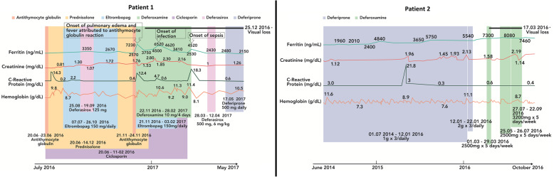
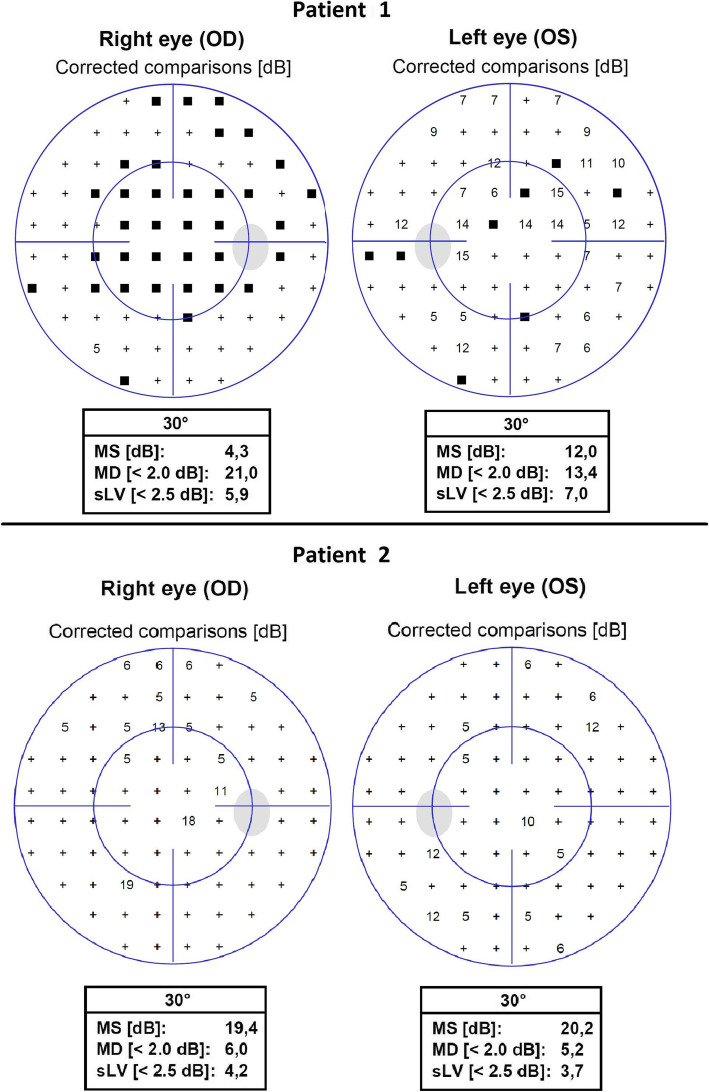
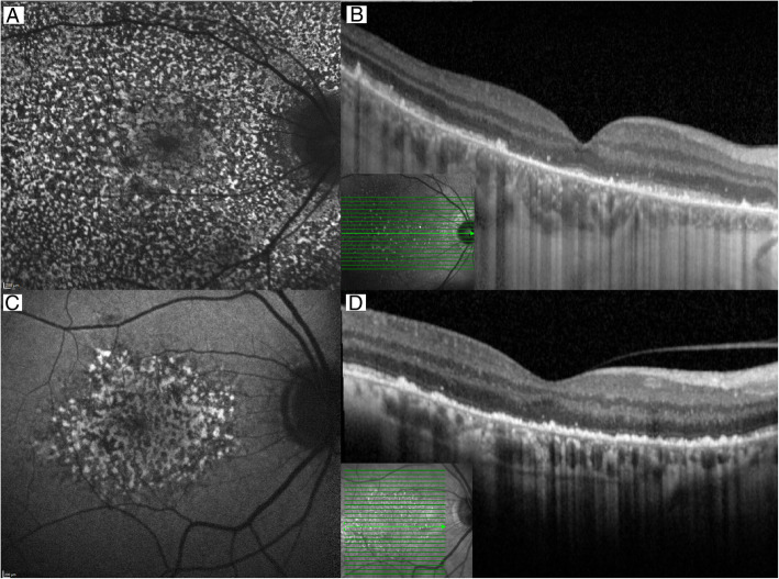
Case presentation: Both anemia patients presented with bilateral visual loss secondary to photoreceptor and retinal pigment epithelium degeneration. Chart review showed that visual loss came after a year-long slow, and rather monotonous rise in plasma ferritin concentrations, with no obvious relation to iron chelator exposure. In one patient, the onset of symptomatic visual loss came after a bout of fever followed by two additional febrile episodes, all accompanied by plasma ferritin spikes. Adjustment of iron chelation therapy did not improve visual function. Experimental studies clearly show that both systemic and intraocular exposure to iron ions can induce retinal degeneration.

Conclusion: The available evidence indicates that retinal degeneration in chronic anemia patients treated by deferoxamine is cause by insufficient iron chelation, not by deferoxamine. The actual role of iron chelating agents may be to promote a long enough survival to allow the slow development of retinal siderosis.

Keywords: Anemia; Case report; Deferoxamine; Iron chelation; Iron overload; Retinopathy.


Conflict of interest statement

ML has consulted, spoken or been a trial investigator for Novartis, Chiesi, Allergan, Bayer, Alcon, AbbVie, Biogen, Novo Nordisk, Eli Lilly, Spark Therapeutics, Nightstar, Sanofi and Roche.

Figures

Fig. 1
Fig. 1
Timeline chart for patient 1 and 2 with relevant blood tests and drug exposure. Legend: Patient 1, timeline from 4 years after presentation with chronic anemia through half a year leading up to the onset of visual loss and 4 months thereafter, showing the exposure to cyclosporin, antithymocyte globulin, iron chelators (deferasirox, deferoxamine and deferiprone), thrombopoietin analog (eltrombopag), onset and persistence of visual loss, onset of fever and pulmonary edema, and plasma concentrations of ferritin, creatinine, c-reactive protein and hemoglobin. Additionally, the patient received blood transfusions throughout the period of observation. Patient 2, timeline from 3 years after the diagnosis of blood dyscrasia through 37 months of follow-up. Deferiprone was exchanges for deferoxamine due to deviating renal function and inefficiency. Additionally, the patient received blood transfusions throughout the period of observation. SI conversion factor: Ferritin, ng/mL ➔ μg/L, multiply by 1; creatinine, mg/dL ➔ μmol/L, multiply by 88.42; c-reactive protein, mg/dL ➔ mg/L, multiply by 10; and hemoglobin, g/dL ➔ mmol/L, multiply by 0.62
Fig. 2
Fig. 2
Automated perimetry examination of patient 1 and 2. Legend: Automated perimetry, 30-2, (Octopus 900, Haag-Streit, Switzerland) in two patients, patient 1 above at presentation and patient 2 below at 2 years after presentation. MS, mean sensitivity; MD, mean defect; and sLV, square root of loss variance
Fig. 3
Fig. 3
Optical coherence tomography and blue light fundus autofluorescence imaging of retinal defects. Legend: In patient 1 (A + B), a diffuse dotted blue light fundus autofluorescence of the right eye shows widespread defects in the whole macula (A). The corresponding horizontal transfoveal optical coherence tomography shows degeneration and irregularities in the outer retina (B). In patient 2 (C + D), a more localized defect is visible in the parafoveal region on blue light fundus autofluorescence (C). The optical coherence tomography shows the same outer retinal defects as in case 1 but temporally an intact outer retina is seen (D). In case 1 the images were acquired during initial visit, and in case 2 the images were acquired at follow-up 6 months after initial visit. Scans were acquired on Spectralis OCT2 (Heidelberg Engineering, Heidelberg, Germany)
Fig. 4
Fig. 4
Adaptive optics fundus photography of photoreceptor mosaic. Legend: Patient 1 (A), patient 2 (B) and a healthy age-matched volunteer (C), spanning from the fovea (indicated by red circles) to a position 14 degrees temporal of the foveal center. Patient 1 is remarkable for having a transitional zone (white arrows) between injured granular retina on the foveal side and more normal retina on the temporal side. Patient 2 shows patchy absence or attenuation of the photoreceptor matrix. The healthy volunteer had a normal photoreceptor distribution. The images of patient 1 and 2 were recorded 5 and 4 months, respectively, after presentation. Acquired on RTX-1 (Imagine Eyes, Orly, France) on flood-illuminated adaptive optics fundus photography

References

    1. Borgna-Pignatti C, Rugolotto S, De Stefano P, et al. Survival and complications in patients with thalassemia major treated with transfusion and deferoxamine. Haematologica. 2004;89(10):1187–1193. - PubMed
    1. Viola F, Barteselli G, DellʼArti L, et al. Multimodal imaging in deferoxamine retinopathy. Retina. 2014;34(7):1428–1438. doi: 10.1097/IAE.0000000000000073. - DOI - PubMed
    1. Davies SC, Marcus RE, Hungerford JL, Miller MH, Arden GB, Huehns ER. Ocular toxicity of high-dose intravenous desferrioxamine. Lancet. 1983;2(8343):181–184. doi: 10.1016/s0140-6736(83)90170-8. - DOI - PubMed
    1. Di Nicola M, Barteselli G, Dell'Arti L, Ratiglia R, Viola F. Functional and structural abnormalities in Deferoxamine retinopathy: a review of the literature. Biomed Res Int. 2015;2015:249617–249612. doi: 10.1155/2015/249617. - DOI - PMC - PubMed
    1. Van Bol L, Alami A, Benghiat FS, Rasquin F. Spectral domain optical coherence tomography findings in early deferoxamine maculopathy: report of two cases. Retinal Cases and Brief Reports. 2014;8(2):97–102. doi: 10.1097/ICB.0000000000000013. - DOI - PubMed

MeSH terms

LinkOut - more resources