

Primary mammary actinomycosis challenged with penicillin allergy
- PMID: 34257106
- PMCID: PMC8278899
- DOI: 10.1136/bcr-2020-235883
Primary mammary actinomycosis challenged with penicillin allergy
Abstract
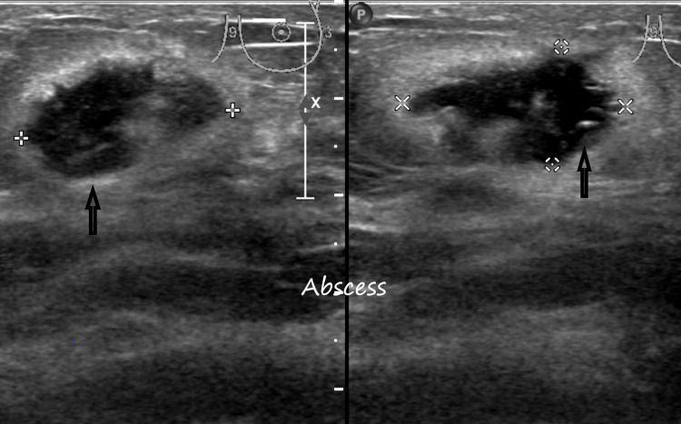
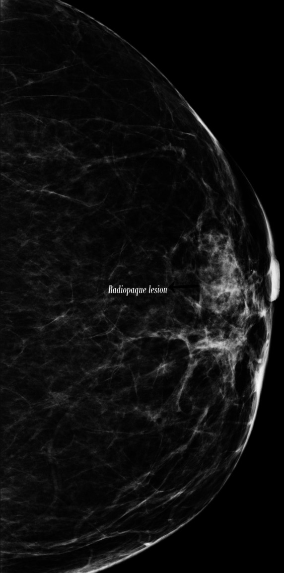
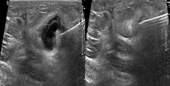
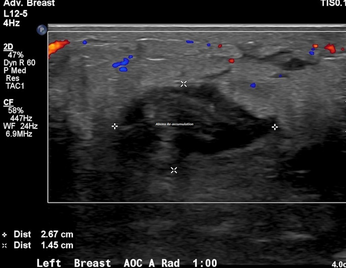
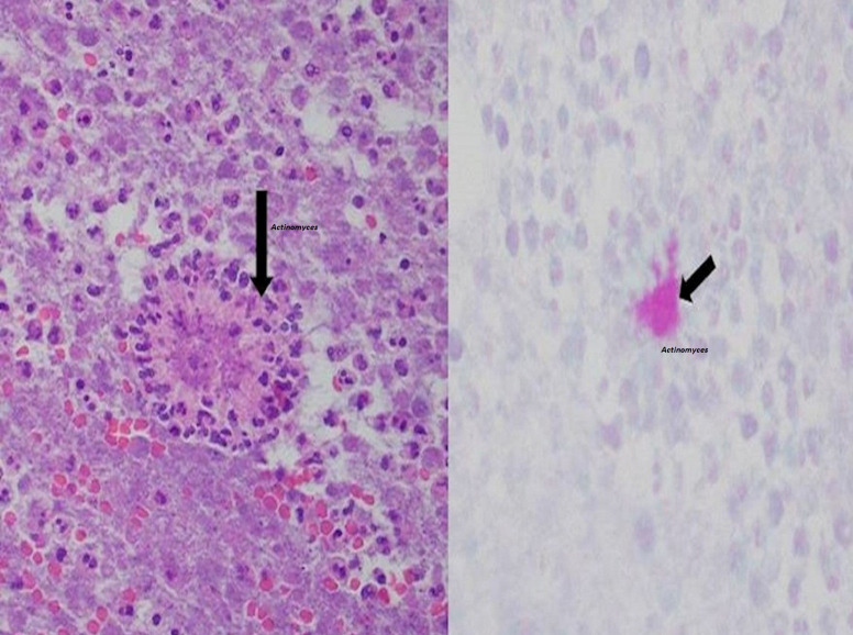
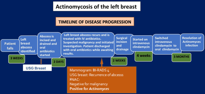
Actinomycosis is a subacute-to-chronic bacterial infection caused by gram-positive, filamentous, non-acid-fast, facultative anaerobic bacteria. It is a normal commensal bacterium found in the oral cavity and the lower reproductive tract of women. We present a case of primary actinomycosis of the breast. A postmenopausal woman, complicated by penicillin allergy, presented with a left breast lump clinically simulating malignancy. The first line of treatment for actinomycosis is penicillin. Due to a penicillin allergy, the patient was initially treated with doxycycline. However, doxycycline was discontinued due to tremors, and was replaced by clindamycin. The patient had a good clinical response with resolution of the abscess.
Keywords: breast cancer; drugs: infectious diseases; general practice / family medicine; general surgery; ultrasonography.
© BMJ Publishing Group Limited 2021. No commercial re-use. See rights and permissions. Published by BMJ.
Conflict of interest statement
Competing interests: None declared.
Figures
Similar articles
-
Treatment of Eikenella corrodens and Actinomyces odontolyticus foot abscess in a penicillin-allergic patient.Ann Pharmacother. 2008 Nov;42(11):1706-10. doi: 10.1345/aph.1L257. Epub 2008 Oct 14. Ann Pharmacother. 2008. PMID: 18854480
-
Adverse Reactions Associated with Penicillins, Carbapenems, Monobactams, and Clindamycin: A Retrospective Population-based Study.J Allergy Clin Immunol Pract. 2020 Apr;8(4):1302-1313.e2. doi: 10.1016/j.jaip.2019.11.035. Epub 2019 Dec 9. J Allergy Clin Immunol Pract. 2020. PMID: 31821919
-
Abdominal actinomycosis.Infection. 2008 Mar;36(2):191. doi: 10.1007/s15010-008-8061-8. Infection. 2008. PMID: 18379724
-
Evaluation and Management of Penicillin Allergy.Mayo Clin Proc. 2018 Jan;93(1):101-107. doi: 10.1016/j.mayocp.2017.09.020. Mayo Clin Proc. 2018. PMID: 29304914 Review.
-
[Actinomycosis of the pelvis with an indwelling IUD].Z Gastroenterol. 2000 May;38(5):375-9. doi: 10.1055/s-2000-14880. Z Gastroenterol. 2000. PMID: 10875147 Review. German.
Cited by
-
Actinomyces spp. Prosthetic Vascular Graft Infection (PVGI): A Multicenter Case-Series and Narrative Review of the Literature.Microorganisms. 2023 Dec 6;11(12):2931. doi: 10.3390/microorganisms11122931. Microorganisms. 2023. PMID: 38138076 Free PMC article. Review.
References
-
- Ghazvini RD, Zaini F, Zibafar E. First case report of primary actinomycosis of the breast due to Actinomyces israelii from Iran. Acta Medica Iranica 2003;41:110–2.
Publication types
MeSH terms
Substances
LinkOut - more resources
Full Text Sources
Medical