. 2021 Jul 13;11(1):14433.
doi: 10.1038/s41598-021-93821-5.

Effects of intracorneal ring segments implementation technique and design on corneal biomechanics and keratometry in a personalized computational analysis

Affiliations

Effects of intracorneal ring segments implementation technique and design on corneal biomechanics and keratometry in a personalized computational analysis

Niksa Mohammadi Bagheri et al. Sci Rep. .

Abstract

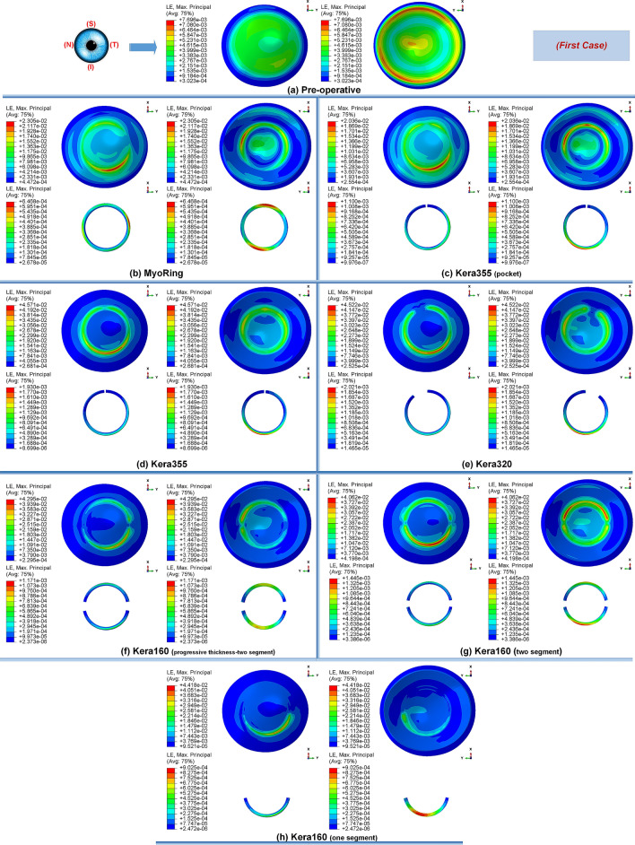
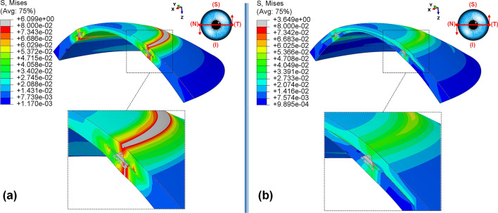
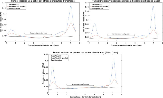
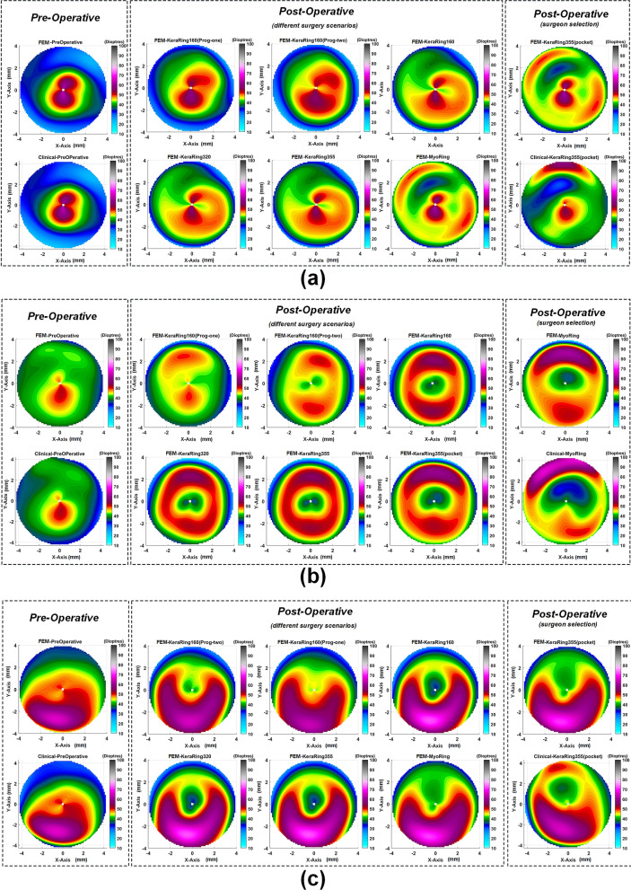
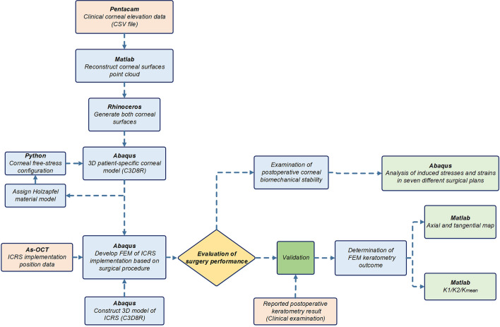
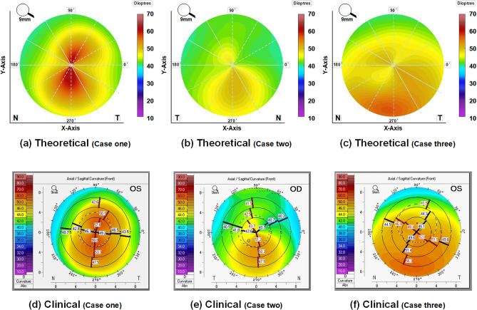
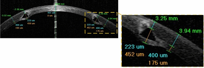
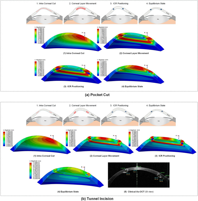
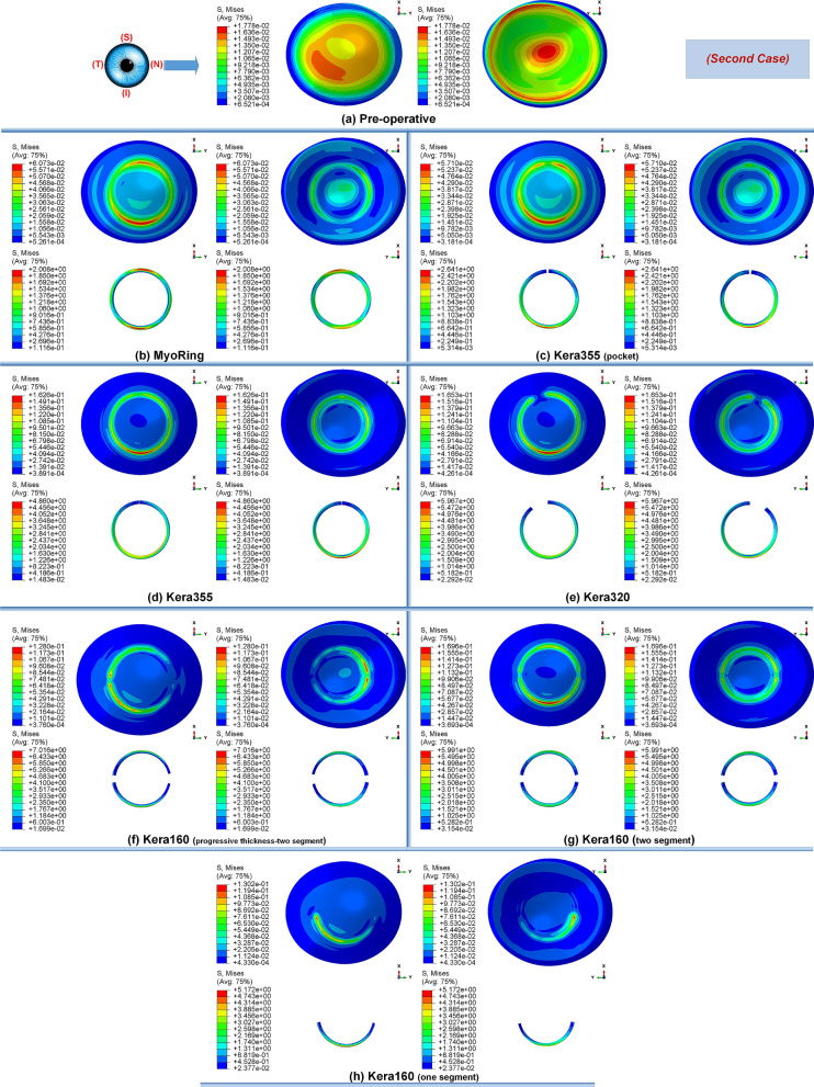
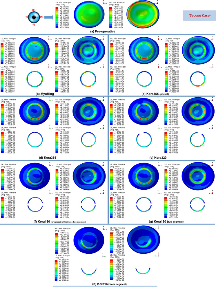
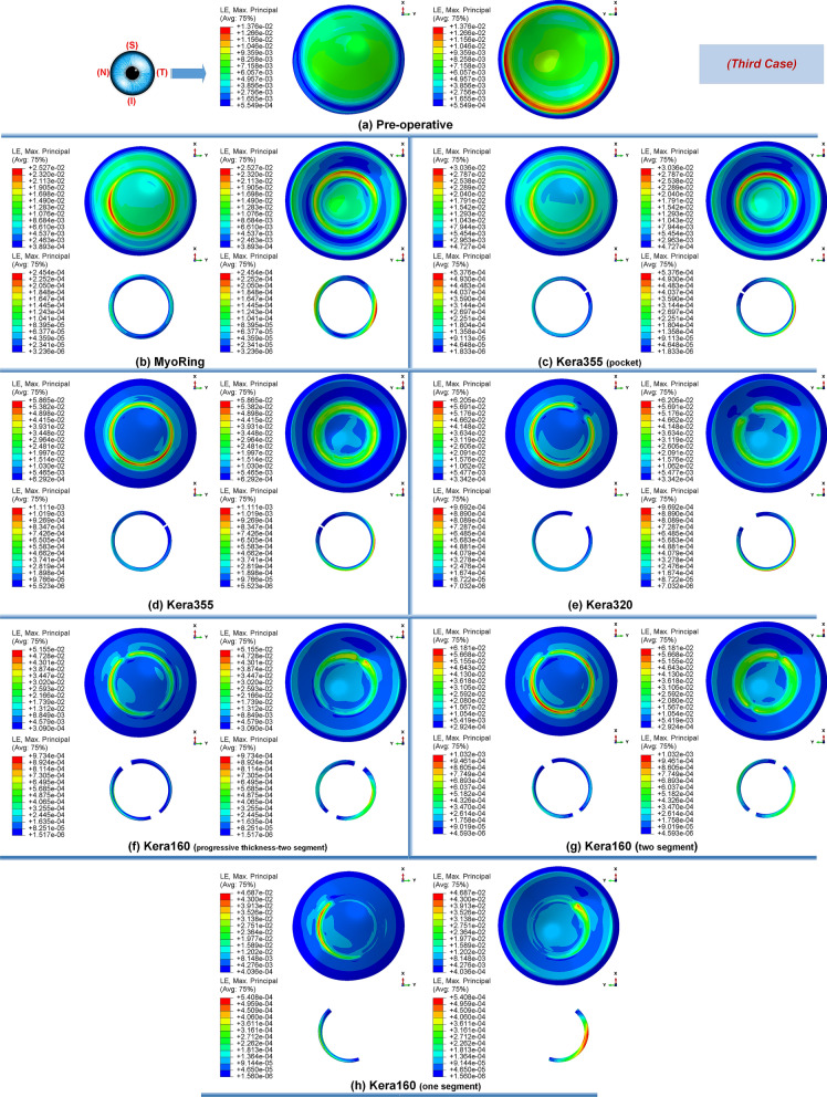
The implementation of intracorneal ring segments (ICRS) is one of the successfully applied refractive operations for the treatment of keratoconus (kc) progression. The different selection of ICRS types along with the surgical implementation techniques can significantly affect surgical outcomes. Thus, this study aimed to investigate the influence of ICRS implementation techniques and design on the postoperative biomechanical state and keratometry results. The clinical data of three patients with different stages and patterns of keratoconus were assessed to develop a three-dimensional (3D) patient-specific finite-element model (FEM) of the keratoconic cornea. For each patient, the exact surgery procedure definitions were interpreted in the step-by-step FEM. Then, seven surgical scenarios, including different ICRS designs (complete and incomplete segment), with two surgical implementation methods (tunnel incision and lamellar pocket cut), were simulated. The pre- and postoperative predicted results of FEM were validated with the corresponding clinical data. For the pre- and postoperative results, the average error of 0.4% and 3.7% for the mean keratometry value ([Formula: see text]) were predicted. Furthermore, the difference in induced flattening effects was negligible for three ICRS types (KeraRing segment with arc-length of 355, 320, and two separate 160) of equal thickness. In contrast, the single and double progressive thickness of KeraRing 160 caused a significantly lower flattening effect compared to the same type with constant thickness. The observations indicated that the greater the segment thickness and arc-length, the lower the induced mean keratometry values. While the application of the tunnel incision method resulted in a lower [Formula: see text] value for moderate and advanced KC, the induced maximum Von Mises stress on the postoperative cornea exceeded the induced maximum stress on the cornea more than two to five times compared to the pocket incision and the preoperative state of the cornea. In particular, an asymmetric regional Von Mises stress on the corneal surface was generated with a progressive ICRS thickness. These findings could be an early biomechanical sign for a later corneal instability and ICRS migration. The developed methodology provided a platform to personalize ICRS refractive surgery with regard to the patient's keratoconus stage in order to facilitate the efficiency and biomechanical stability of the surgery.


Conflict of interest statement

The authors declare no competing interests.

Figures

Figure 1
Figure 1
The pre- and postoperative contour diagrams of the Von Mises stress distribution (MPa) in FEM of different surgical scenarios (first case): (a) preoperative; (b) MyoRing; (c) Kera355 implemented by pocket incision; (d) Kera355; (e) Kera320; (f) Kera160-two prog segments; (g) Kera160-two segments; (h) Kera160-one prog segment.
Figure 2
Figure 2
The pre- and postoperative contour plots of the principal strain’s distribution in FEM of the different surgical scenarios (first case): (a) preoperative; (b) MyoRing; (c) Kera355 implemented by pocket incision; (d) Kera355; (e) Kera320; (f) Kera160-two prog segments; (g) Kera160-two segments; (h) Kera160-one prog segment.
Figure 3
Figure 3
The postoperative contour of the Von Mises stress distribution (MPa) in the nasal-temporal cross-section of FEM of Kera355 implantation using (a) tunnel incision; (b) lamellar pocket cut (case one).
Figure 4
Figure 4
2D comparison plot of Von Mises stress distribution in superior-inferior direction on the anterior corneal surface for all case scenario. (Kera355 implemented by tunnel incision and pocket incision are shown in the figure by light blue (dashed-line) and red (dotted-line) respectively).
Figure 5
Figure 5
Comparison of the Von Mises stress distribution (MPa) and the contour of locally induced stresses at the ICRS endpoints for three different types of ICRS, including: (a) Kera160-two prog segments; (b) Kera160-two segments; (c) Kera160-one prog segment.
Figure 6
Figure 6
The pre- and postoperative maps of tangential curvature tomography (D) obtained at FEM from different surgical scenarios with respect to clinical reported maps for the (a) first case, (b) second case, (c) third case.
Figure 7
Figure 7
The schematic flowchart of the steps used in the development and evaluation of the patient-specific FEM of ICRS implementation surgery.
Figure 8
Figure 8
The preoperative axial maps: theoretically obtained from Pentacam elevation data for three individual case scenarios (the first row); the clinically reported axial maps for each case (the second row).
Figure 9
Figure 9
(a) The patient-specific 3D construction of the cornea FEM. The defined variables of developed ICRS in 3D FEM (b) KeraRing, (c) MyoRing, (d) progressive KeraRing.
Figure 10
Figure 10
The postoperative AS-OCT image of ICRS implementation surgery (case three).
Figure 11
Figure 11
The summary of the step-by-step ICRS implementation methods; (a) pocket cut; (b) tunnel incision.
Figure 12
Figure 12
The pre- and post-operative contour diagrams of the Von Mises stress distribution (MPa) in FEM of different surgical scenarios (second case): (a) preoperative; (b) MyoRing; (c) Kera355 implemented by pocket incision; (d) Kera355; (e) Kera320; (f) Kera160-two prog segments; (g) Kera160-two segments; (h) Kera160-one prog segment.
Figure 13
Figure 13
The pre- and postoperative contour plots of the principal strain’s distribution in FEM of the different surgical scenarios (second case): (a) Preoperative; (b) MyoRing; (c) Kera355 implemented by pocket incision; (d) Kera355; (e) Kera320; (f) Kera160-two prog segments; (g) Kera160-two segments; (h) Kera160-one prog segment.
Figure 14
Figure 14
The pre- and post-operative contour diagrams of the Von Mises stress distribution (MPa) in FEM of different surgical scenarios (third case): (a) preoperative; (b) MyoRing; (c) Kera355 implemented by pocket incision; (d) Kera355; (e) Kera320; (f) Kera160-two prog segments; (g) Kera160-two segments; (h) Kera160-one prog segment.
Figure 15
Figure 15
The pre- and postoperative contour plots of the principal strain’s distribution in FEM of the different surgical scenarios (third case): (a) Preoperative; (b) MyoRing; (c) Kera355 implemented by pocket incision; (d) Kera355; (e) Kera320; (f) Kera160-two prog segments; (g) Kera160-two segments; (h) Kera160-one prog segment.

References

    1. Auffarth GU, Wang L, Völcker HE. Keratoconus evaluation using the orbscan topography system. J. Cataract Refract. Surg. 2000;26:222–228. doi: 10.1016/S0886-3350(99)00355-7. - DOI - PubMed
    1. Vega-Estrada A, Alió JL, Puche ABP, Marshall J. Outcomes of a new microwave procedure followed by accelerated cross-linking for the treatment of keratoconus: A pilot study. J. Refract. Surg. 2012;28:787–793. doi: 10.3928/1081597X-20121011-07. - DOI - PubMed
    1. Vega-Estrada A, et al. Outcome analysis of intracorneal ring segments for the treatment of keratoconus based on visual, refractive, and aberrometric impairment. Am. J. Ophthalmol. 2013;155:575–584.e1. doi: 10.1016/j.ajo.2012.08.020. - DOI - PubMed
    1. Watson SL, Ramsay A, Dart JK, Bunce C, Craig E. Comparison of deep lamellar keratoplasty and penetrating keratoplasty in patients with keratoconus. Ophthalmology. 2004;111:1676–1682. doi: 10.1016/j.ophtha.2004.02.010. - DOI - PubMed
    1. d’Azy CB, Pereira B, Chiambaretta F, Dutheil F. Efficacy of different procedures of intra-corneal ring segment implantation in keratoconus: A systematic review and meta-analysis. Transl. Vis. Sci. Technol. 2019;8:38. doi: 10.1167/tvst.8.3.38. - DOI - PMC - PubMed