

CAD systems for colorectal cancer from WSI are still not ready for clinical acceptance
- PMID: 34257363
- PMCID: PMC8277780
- DOI: 10.1038/s41598-021-93746-z
CAD systems for colorectal cancer from WSI are still not ready for clinical acceptance
Abstract
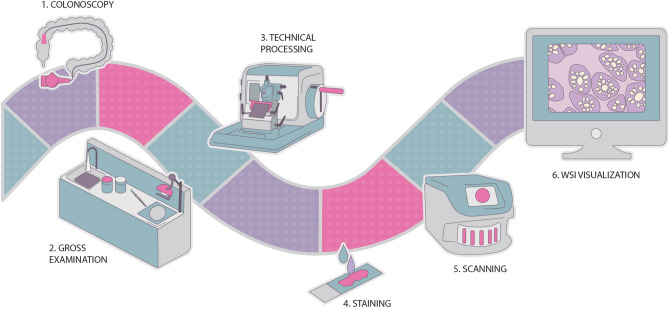
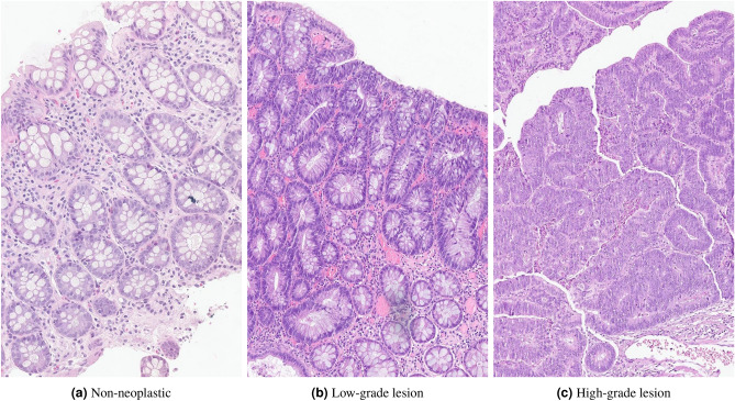
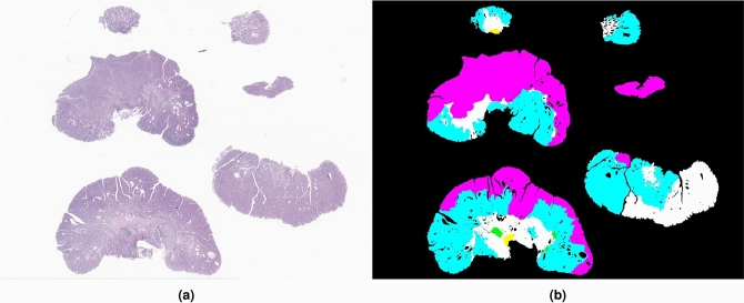
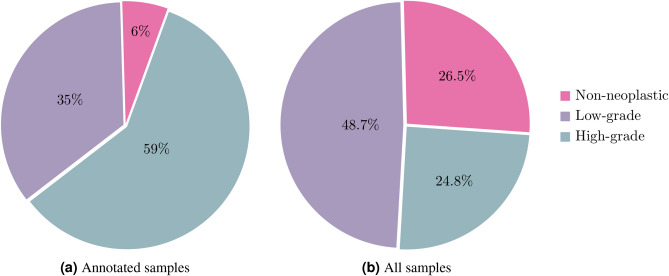
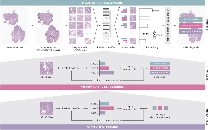
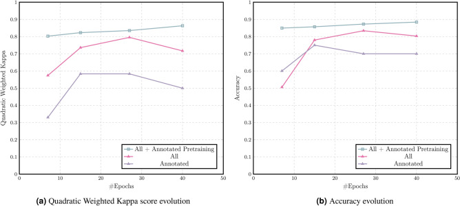
Most oncological cases can be detected by imaging techniques, but diagnosis is based on pathological assessment of tissue samples. In recent years, the pathology field has evolved to a digital era where tissue samples are digitised and evaluated on screen. As a result, digital pathology opened up many research opportunities, allowing the development of more advanced image processing techniques, as well as artificial intelligence (AI) methodologies. Nevertheless, despite colorectal cancer (CRC) being the second deadliest cancer type worldwide, with increasing incidence rates, the application of AI for CRC diagnosis, particularly on whole-slide images (WSI), is still a young field. In this review, we analyse some relevant works published on this particular task and highlight the limitations that hinder the application of these works in clinical practice. We also empirically investigate the feasibility of using weakly annotated datasets to support the development of computer-aided diagnosis systems for CRC from WSI. Our study underscores the need for large datasets in this field and the use of an appropriate learning methodology to gain the most benefit from partially annotated datasets. The CRC WSI dataset used in this study, containing 1,133 colorectal biopsy and polypectomy samples, is available upon reasonable request.
© 2021. The Author(s).
Conflict of interest statement
The authors declare no competing interests.
Figures
References
Publication types
MeSH terms
LinkOut - more resources
Full Text Sources
Medical
Research Materials
Miscellaneous
