

Osteoclasts in Tumor Biology: Metastasis and Epithelial-Mesenchymal-Myeloid Transition
- PMID: 34257573
- PMCID: PMC8262221
- DOI: 10.3389/pore.2021.609472
Osteoclasts in Tumor Biology: Metastasis and Epithelial-Mesenchymal-Myeloid Transition
Abstract
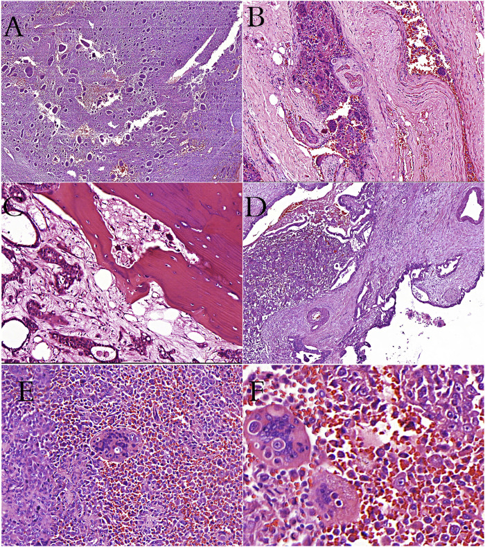
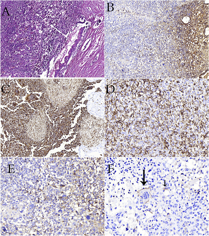
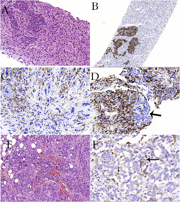
Osteoclast is a specialized cell that originates from monocytic lineage, communicates closely with osteoblasts under physiological conditions, participates in bone modeling and re-modeling, contributes to calcium homeostasis and osteoimmunity. In pathological conditions, it is involved in many tumors such as giant cell bone tumor (osteoclastoma), aneurysmal bone cyst, osteosarcoma, and metastatic cancers, and it usually causes local spread and progression of the tumor, working against the host. Since osteoclasts play an active role in primary bone tumors and bone metastases, the use of anti-osteoclastic agents significantly reduces the mortality and morbidity rates of patients by preventing the progression and local spread of tumors. Osteoclasts also accompany undifferentiated carcinomas of many organs, especially pancreas, thyroid, bladder and ovary. Undifferentiated carcinomas rich in osteoclasts have osteoclastoma-like histology. In these organs, osteoclastoma-like histology may accompany epithelial carcinomas, and de novo, benign and borderline tumors. Mature and immature myeloid cells, including osteoclasts, play an active role in the tumor progression in primary and metastatic tumor microenvironment, in epithelial-mesenchymal transition (EMT), mesenchymal-epithelial-transition (MET), and cancer stem cell formation. Additionally, they are the most suitable candidates for cancer cells in cell fusion due to their evolutionary fusion capabilities. Myeloid features and markers (CD163, CD33, CD68 etc.) can be seen in metastatic cancer cells. Consequently, they provide metastatic cancer cells with motility, margination, transmigration, chemotaxis, phagocytosis, angiogenesis, matrix degradation, and resistance to chemotherapy. For these reasons, we think that the concept of Epithelial-Mesencyhmal-Myeloid-Transition (EMMT) will be more accurate than EMT for cancer cells with myeloid properties.
Keywords: EMMT; breast carcinoma metastasis; epithelial-mesenchymal-myeloid transition; giant cell tumor of bone; metastasis; osteoclast; tumor biology.
Copyright © 2021 Behzatoglu.
Conflict of interest statement
The author declares that the research was conducted in the absence of any commercial or financial relationships that could be construed as a potential conflict of interest.
Figures
References
-
- Scully OJ, Bay BH, Yip G, Yu Y. Breast cancer metastasis. Cancer Genomics Proteomics (2012) 9(5):311–20. - PubMed
Publication types
MeSH terms
LinkOut - more resources
Full Text Sources
Medical
Research Materials
Miscellaneous