. 2021 May 8;8(Pt 4):574-583.
doi: 10.1107/S2052252521003729. eCollection 2021 Jul 1.

Putative hexameric glycosyltransferase functional unit revealed by the crystal structure of Acinetobacter baumannii MurG

Affiliations

Putative hexameric glycosyltransferase functional unit revealed by the crystal structure of Acinetobacter baumannii MurG

Kyoung Ho Jung et al. IUCrJ. .

Abstract

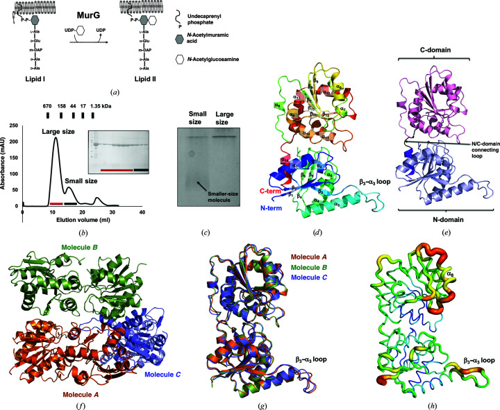
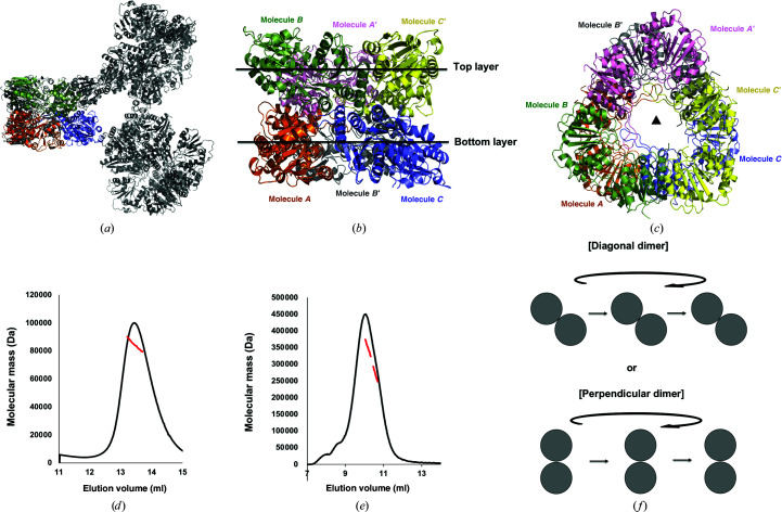
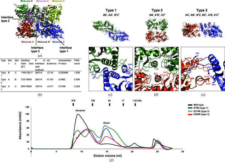
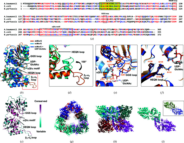
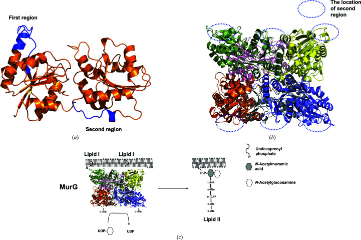
Lipid II, the main component of the bacterial cell wall, is synthesized by the addition of UDP-N-acetylglucosamine to the UDP-N-acetylmuramic acid pentapeptide catalyzed by the glycosyltransferase MurG. Owing to its critical role in cell-wall biosynthesis, MurG is considered to be an attractive target for antibacterial agents. Although the Mur family ligases have been extensively studied, the molecular mechanism of the oligomeric scaffolding assembly of MurG remains unclear. In this study, MurG from Acinetobacter baumannii (abMurG), a human pathogen, was characterized and its hexameric crystal structure was unveiled; this is the first homo-oligomeric structure to be described in the MurG family and the Mur family. Homogeneous protein samples were produced for structural studies using size-exclusion chromatography, the absolute molecular mass was calculated via multi-angle light scattering, and protein-protein interactions were analyzed using the PDBePISA server. abMurG was found to form homo-oligomeric complexes in solution, which might serve as functional units for the scaffolding activity of MurG. Furthermore, analysis of this structure revealed the molecular assembly mechanism of MurG. This structural and biochemical study elucidated the homo-oligomerization mechanism of MurG and suggests a new potential antibiotic target on MurG.

Keywords: Acinetobacter baumannii; MurG; cell-wall peptidoglycan biosynthesis; crystal structure; glycosyltransferases; superbugs.


Figures

Figure 1
Figure 1
Crystal structure of abMurG. (a) Overview of the function of MurG. (b) Size-exclusion chromatography profile. Two peaks are labeled corresponding to an oligomer and a dimer. SDS–PAGE to assess the identity and purity is shown to the right of the two main peaks. The loaded fractions are indicated by black and red bars. (c) Native PAGE gel. The loaded samples are indicated above the gel. (d) Multi-colored cartoon representation of monomeric abMurG. The chain from the N- to C-terminus is colored from blue to red. Helices and sheets are labeled α and β, respectively. (e) A cartoon representation of the structure of abMurG showing the domain boundary in the structure. (f) Cartoon representation of the three abMurG molecules in an asymmetric unit. (g) Superposition of the structures of the molecules found in one asymmetric unit. (h) Putty representation showing the B-factor distribution. Rainbow colors from red to violet with increasing B-factor values were used for B-factor visualization.
Figure 2
Figure 2
Hexameric structure of abMurG. (a) Crystallographic packing symmetry analysis. Three abMurG molecules in the asymmetric unit are indicated by color cartoons, whereas other symmetric molecules are indicated by gray ribbon structures. (b, c) Tentative hexameric structure of abMurG generated by the symmetry analysis: side (b) and top (c) views. (d, e) Multi-angle light scattering (MALS) profiles derived from the first (d) and second (e) size-exclusion chromatography peaks. The red line indicates the experimental molecular mass analyzed by MALS. (f) Schematic planar diagram showing the hexamer assembly strategy of two tentative dimers.
Figure 3
Figure 3
Details of the interface formed by the hexameric homo-oligomeric complex of abMurG. (a) Cartoon representation of the hexameric abMurG complex. Three different types of interfaces (types 1–3) formed by the hexameric complex are indicated by black arrows. (b) Table summarizing the interaction details of each type of interface. (c, d, e) Cartoon representation of each type of interaction interface: type 1 (c), type 2 (d) and type 3 (e). The position of each interface and the molecules involved in the formation of each interface are shown in the upper panel. Close-up views of each interface showing the residues involved in the formation of the interface are provided in the lower panels. (f) Verification of the interfaces by mutagenesis. Size-exclusion chromatography profiles comparing the positions of eluted peaks between wild-type abMurG and various mutants with disrupted type 1 (R76D), type 2 (S319K) and type 3 (E209R) interfaces.
Figure 4
Figure 4
Structure and sequence comparison of abMurG and ecMurG. (a) Sequence alignment of MurGs from different species. Mostly conserved and partially conserved residues are shown in red and blue, respectively. The HEQN loop, GGS loop and α/β/α motif are indicated with black boxes. The position of the β3–α3 loop is highlighted. (b) Structural superposition of abMurG (orange) with ecMurG (cyan) and the ecMurG–UDP–GlcNAc complex (blue). The red dotted box indicates the β3–α3 loop region. (c) Cartoon representation of abMurG colored according to the degree of amino-acid sequence conservation. The HEQN loop, GGS loop and α/β/α motif regions, which are critical for the accommodation of substrates in the structure of abMurG, are indicated. (d) Different structural details of the β3–α3 loop region between abMurG (orange) and ecMurG (cyan). (e) Superimposition of the structure of abMurG with ecMurG focusing on the UDP–GlcNAc-binding site. (f) Superimposition of the structure of abMurG with ecMurG focusing on the HEQN loop. (g, h) Putative hexameric structure of ecMurG modeled using the hexameric abMurG structure as a template: top (g) and side (h) views. (i) Type 1 interaction between two molecules in the hexameric structure of ecMurG. The type 1 interaction in the hexameric structure of abMurG is shown in a black box for comparison.
Figure 5
Figure 5
Proposed working model of hexameric MurG on the membrane. (a) Tentative membrane-docking regions on MurG. Membrane-anchoring regions were predicted by the MODA server. Two regions, colored blue, were analyzed as tentative membrane-docking regions. (b) The location of the second region, formed by a small helix connected by a loop, on each subunit of hexameric MurG. (c) Tentative working model of hexameric MurG on the membrane.

References

    1. Basavannacharya, C., Moody, P. R., Munshi, T., Cronin, N., Keep, N. H. & Bhakta, S. (2010). Protein Cell, 1, 1011–1022. - PMC - PubMed
    1. Brink-van der Laan, E. van den, Boots, J. P., Spelbrink, R. E. J., Kool, G. M., Breukink, E., Killian, J. A. & de Kruijff, B. (2003). J. Bacteriol. 185, 3773–3779. - PMC - PubMed
    1. Bupp, K. & van Heijenoort, J. (1993). J. Bacteriol. 175, 1841–1843. - PMC - PubMed
    1. Burki, T. K. (2018). Lancet Respir. Med. 6, 668. - PubMed
    1. Chen, V. B., Arendall, W. B., Headd, J. J., Keedy, D. A., Immormino, R. M., Kapral, G. J., Murray, L. W., Richardson, J. S. & Richardson, D. C. (2010). Acta Cryst. D66, 12–21. - PMC - PubMed