

Mechanistic Targets of Diallyl Trisulfide in Human Breast Cancer Cells Identified by RNA-seq Analysis
- PMID: 34258251
- PMCID: PMC8249207
- DOI: 10.15430/JCP.2021.26.2.128
Mechanistic Targets of Diallyl Trisulfide in Human Breast Cancer Cells Identified by RNA-seq Analysis
Abstract
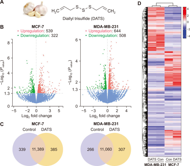
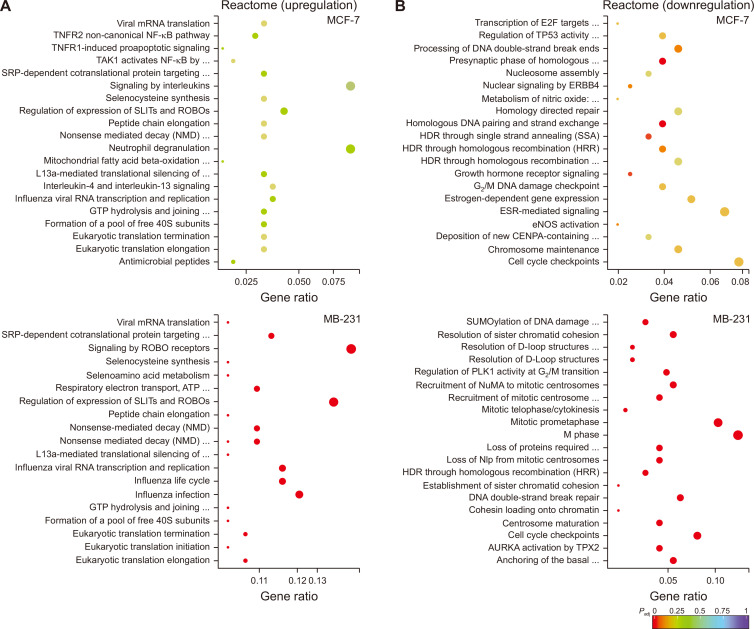
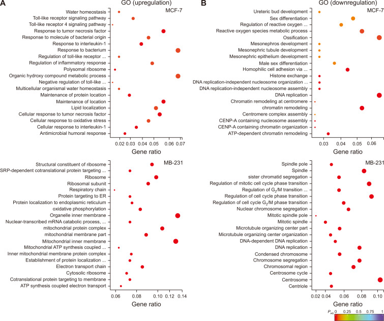
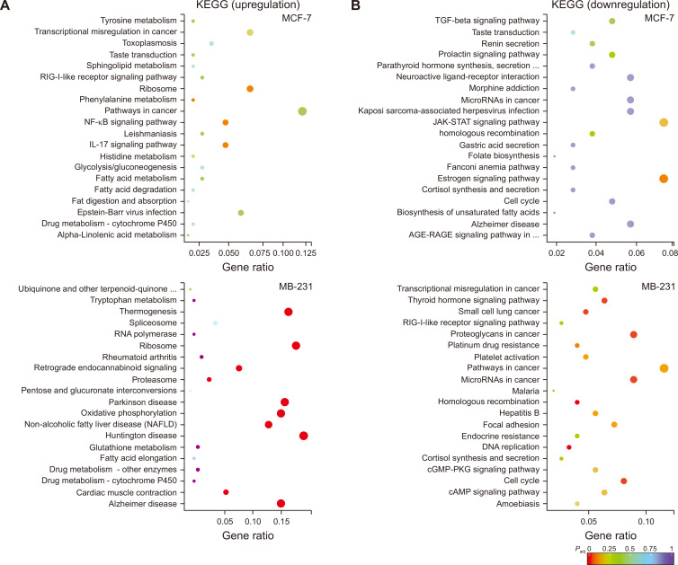
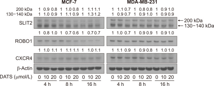
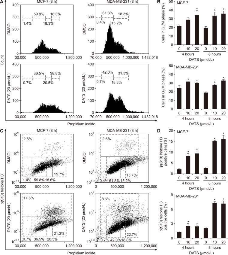
Diallyl trisulfide (DATS), a metabolic by-product of processed garlic, is highly effective in inhibiting growth of human breast cancer cells in vitro and in vivo, but the underlying mechanisms are still not fully understood. In this study, we performed RNA-seq analyses using luminal-type (MCF-7) and basal-like (MDA-MB-231) human breast cancer cells to identify mechanistic targets of DATS. The Reactome Pathway Analysis revealed upregulation of genes associated with SLIT/ROBO tumor suppressor signaling following DATS treatment in both MCF-7 and MDA-MB-231 cells. However, the expression of SLIT2 and ROBO1 proteins or their downstream target C-X-C motif chemokine receptor 4 was not affected by DATS treatment in both cell lines. The Reactome as well as the Gene Ontology Pathways Analyses of the RNA-seq data from DATS-treated cells indicated downregulation of genes associated with G2/M phase cell cycle arrest in comparison with vehicle-treated control cells. Consistent with the RNA-seq data, DATS treatment caused a significant increase in the fraction of the G2/M population in both cell lines when compared to corresponding control cells. In addition, Ser10 phosphorylation of histone H3, a mitotic marker, was also increased significantly following DATS treatment in MCF-7 and MDA-MB-231 cells. These results indicate that while SLIT/ROBO signaling is not affected by DATS treatment, cell cycle arrest likely contributes to the antitumor effect of this phytochemical.
Keywords: Allyl compounds; Breast neoplasms; Chemoprevention; Sulfides.
Copyright © 2021 Korean Society of Cancer Prevention.
Conflict of interest statement
CONFLICTS OF INTEREST No potential conflicts of interest were disclosed.
Figures
Similar articles
-
Breast Cancer Selective Disruption of Actin Cytoskeleton by Diallyl Trisulfide.J Cancer Prev. 2022 Jun 30;27(2):101-111. doi: 10.15430/JCP.2022.27.2.101. J Cancer Prev. 2022. PMID: 35864856 Free PMC article.
-
Critical role for reactive oxygen species in apoptosis induction and cell migration inhibition by diallyl trisulfide, a cancer chemopreventive component of garlic.Breast Cancer Res Treat. 2013 Feb;138(1):69-79. doi: 10.1007/s10549-013-2440-2. Epub 2013 Feb 15. Breast Cancer Res Treat. 2013. PMID: 23412769 Free PMC article.
-
Insight into drug sensitizing effect of diallyl disulfide and diallyl trisulfide from Allium sativum L. on paclitaxel-resistant triple-negative breast cancer cells.J Ethnopharmacol. 2022 Oct 5;296:115452. doi: 10.1016/j.jep.2022.115452. Epub 2022 Jun 8. J Ethnopharmacol. 2022. PMID: 35690339
-
Attenuative Effect of Diallyl Trisulfide on Caspase Activity in TNF-α-induced Triple Negative Breast Cancer Cells.Anticancer Res. 2023 Jun;43(6):2393-2405. doi: 10.21873/anticanres.16407. Anticancer Res. 2023. PMID: 37247921 Free PMC article.
-
Dietary Bioactive Diallyl Trisulfide in Cancer Prevention and Treatment.Int J Mol Sci. 2017 Jul 28;18(8):1645. doi: 10.3390/ijms18081645. Int J Mol Sci. 2017. PMID: 28788092 Free PMC article. Review.
Cited by
-
Novel estrogen-responsive genes (ERGs) for the evaluation of estrogenic activity.PLoS One. 2022 Aug 17;17(8):e0273164. doi: 10.1371/journal.pone.0273164. eCollection 2022. PLoS One. 2022. PMID: 35976950 Free PMC article.
-
Diallyl Trisulfide Induces ROS-Mediated Mitotic Arrest and Apoptosis and Inhibits HNSCC Tumor Growth and Cancer Stemness.Cancers (Basel). 2024 Jan 16;16(2):378. doi: 10.3390/cancers16020378. Cancers (Basel). 2024. PMID: 38254868 Free PMC article.
-
Updates on the anticancer potential of garlic organosulfur compounds and their nanoformulations: Plant therapeutics in cancer management.Front Pharmacol. 2023 Mar 20;14:1154034. doi: 10.3389/fphar.2023.1154034. eCollection 2023. Front Pharmacol. 2023. PMID: 37021043 Free PMC article. Review.
-
Cancer Stem Cells Connecting to Immunotherapy: Key Insights, Challenges, and Potential Treatment Opportunities.Cancers (Basel). 2025 Jun 23;17(13):2100. doi: 10.3390/cancers17132100. Cancers (Basel). 2025. PMID: 40647402 Free PMC article. Review.
-
Breast Cancer Selective Disruption of Actin Cytoskeleton by Diallyl Trisulfide.J Cancer Prev. 2022 Jun 30;27(2):101-111. doi: 10.15430/JCP.2022.27.2.101. J Cancer Prev. 2022. PMID: 35864856 Free PMC article.
References
Grants and funding
LinkOut - more resources
Full Text Sources
Molecular Biology Databases
Miscellaneous