

Short-Term Increased Physical Activity During Early Life Affects High-Fat Diet-Induced Bone Loss in Young Adult Mice
- PMID: 34258504
- PMCID: PMC8260814
- DOI: 10.1002/jbm4.10508
Short-Term Increased Physical Activity During Early Life Affects High-Fat Diet-Induced Bone Loss in Young Adult Mice
Abstract
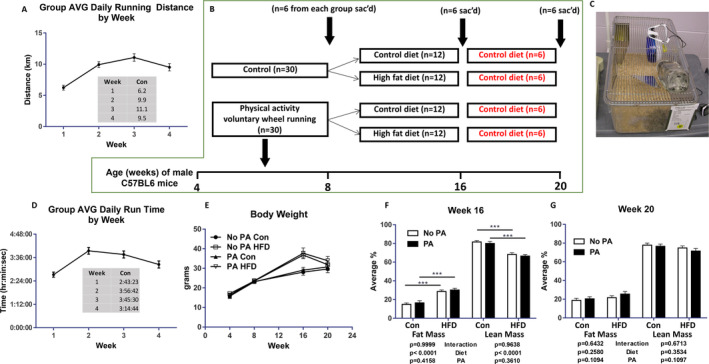
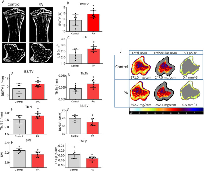
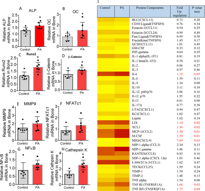
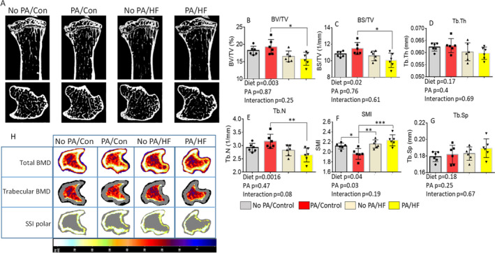
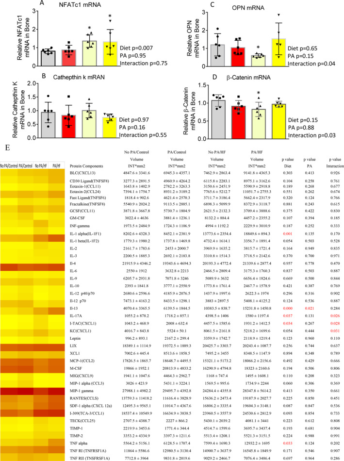
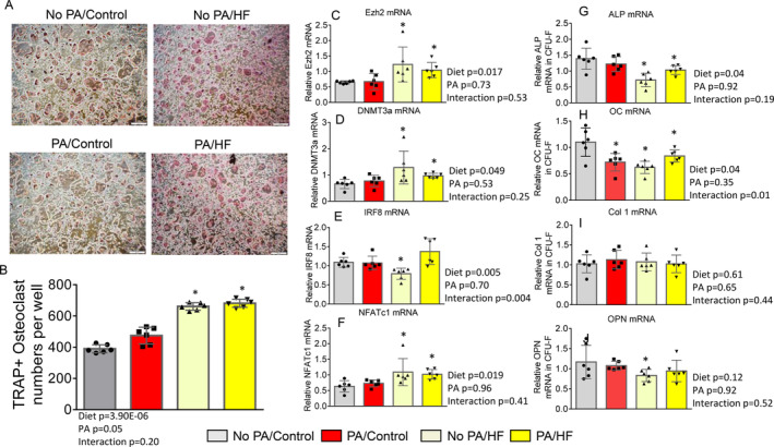
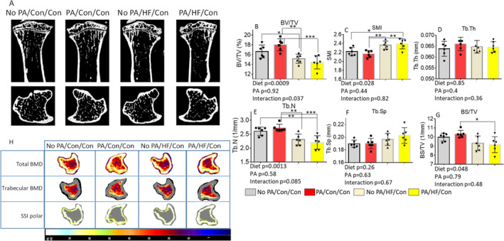
Mechanical stresses associated with physical activity (PA) have beneficial effects on increasing BMD and improving bone quality. However, a high-fat diet (HFD) and obesity tend to have negative effects on bone, by increasing bone marrow adiposity leading to increased excretion of proinflammatory cytokines, which activate RANKL-induced bone resorption. In the current study, whether short-term increased PA via access to voluntary wheel running during early life has persistent and protective effects on HFD-induced bone resorption was investigated. Sixty 4-week-old male C57BL6/J mice were divided into two groups postweaning: without or with PA (access to voluntary running wheel 7-8 km/day) for 4 weeks. After 4 weeks with or without PA, mice were further subdivided into control diet or HFD groups for 8 weeks, and then all animals were switched back to control diet for an additional 4 weeks. Mice from the HFD groups were significantly heavier and obese; however, after 4 weeks of additional control diet their body weights returned to levels of mice on continuous control diet. Using μ-CT and confirmed by pQCT of tibias and spines ex vivo, it was determined that bone volume and trabecular BMD were significantly increased with PA in control diet animals compared with sedentary animals without access to wheels, and such anabolic effects of PA on bone were sustained after ceasing PA in adult mice. Eight weeks of a HFD deteriorated bone development in mice. Unexpectedly, early-life PA did not prevent persistent effects of HFD on deteriorating bone quality; in fact, it exacerbated a HFD-induced inflammation, osteoclastogenesis, and trabecular bone loss in adult mice. In accordance with these data, signal transduction studies revealed that a HFD-induced Ezh2, DNA methyltransferase 3a, and nuclear factor of activated T-cells 1 expression were amplified in nonadherent hematopoietic cells. In conclusion, short-term increased PA in early life is capable of increasing bone mass; however, it alters the HFD-induced bone marrow hematopoietic cell-differentiation program to exacerbate increased bone resorption if PA is halted. © 2021 Arkansas Children's Nutrition Center. JBMR Plus published by Wiley Periodicals LLC on behalf of American Society for Bone and Mineral Research.
Keywords: BONE RESORPTION; EXERCISE; HIGH‐FAT DIET; WHEEL RUNNING.
© 2021 Arkansas Children's Nutrition Center. JBMR Plus published by Wiley Periodicals LLC on behalf of American Society for Bone and Mineral Research.
Figures
Similar articles
-
Voluntary running of defined distances alters bone microstructure in C57BL/6 mice fed a high-fat diet.Appl Physiol Nutr Metab. 2021 Nov;46(11):1337-1344. doi: 10.1139/apnm-2021-0061. Epub 2021 May 17. Appl Physiol Nutr Metab. 2021. PMID: 34000207
-
Deletion of Rptor in Preosteoblasts Reveals a Role for the Mammalian Target of Rapamycin Complex 1 (mTORC1) Complex in Dietary-Induced Changes to Bone Mass and Glucose Homeostasis in Female Mice.JBMR Plus. 2021 Mar 24;5(5):e10486. doi: 10.1002/jbm4.10486. eCollection 2021 May. JBMR Plus. 2021. PMID: 33977204 Free PMC article.
-
Changes in Skeletal Integrity and Marrow Adiposity during High-Fat Diet and after Weight Loss.Front Endocrinol (Lausanne). 2016 Jul 27;7:102. doi: 10.3389/fendo.2016.00102. eCollection 2016. Front Endocrinol (Lausanne). 2016. PMID: 27512386 Free PMC article.
-
A high-fat diet induces obesity and impairs bone acquisition in young male mice.Mol Med Rep. 2013 Apr;7(4):1203-8. doi: 10.3892/mmr.2013.1297. Epub 2013 Jan 29. Mol Med Rep. 2013. PMID: 23444006
-
Contributions of Dickkopf-1 to Obesity-Induced Bone Loss and Marrow Adiposity.JBMR Plus. 2020 Apr 28;4(6):e10364. doi: 10.1002/jbm4.10364. eCollection 2020 Jun. JBMR Plus. 2020. PMID: 32537550 Free PMC article.
Cited by
-
Wheel-Running Exercise Protects Ovariectomized Mice from Bone Loss via IFN-γ-Mediated Suppression of the NF-κB and MAPK Pathways.Oxid Med Cell Longev. 2022 May 11;2022:2030818. doi: 10.1155/2022/2030818. eCollection 2022. Oxid Med Cell Longev. 2022. PMID: 35602099 Free PMC article.
-
Association of a Body Shape Index with Bone Mineral Density and Osteoporosis Among U.S. Adults: Evidence from NHANES.Calcif Tissue Int. 2025 May 25;116(1):76. doi: 10.1007/s00223-025-01386-6. Calcif Tissue Int. 2025. PMID: 40415018
References
-
- Rogers RS, Hinton PS. Bone loading during young adulthood predicts bone mineral density in physically active, middle‐aged men. Phys Sportsmed. 2010;38:146‐155. - PubMed
-
- Baxter‐Jones AD, Kontulainen SA, Faulkner RA, Bailey DA. A longitudinal study of the relationship of physical activity to bone mineral accrual from adolescence to young adulthood. Bone. 2008;43:1101‐1107. - PubMed
-
- Nilsson M, Ohlsson C, Mellström D, Lorentzon M. Previous sport activity during childhood and adolescence is associated with increased cortical bone size in young adult men. J Bone Miner Res. 2009;24:125‐133. - PubMed
-
- Van Langendonck L, Lefevre J, Claessens AL, et al. Influence of participation in high‐impact sports during adolescence and adulthood on bone mineral density in middle‐aged men: a 27‐year follow‐up study. Am J Epidemiol. 2003;158:525‐533. - PubMed
Grants and funding
LinkOut - more resources
Full Text Sources
Research Materials