. 2021 Jul 14;7(1):67.
doi: 10.1186/s40729-021-00347-6.

Impact of the blooming artefact on dental implant dimensions in 13 cone-beam computed tomography devices

Affiliations

Impact of the blooming artefact on dental implant dimensions in 13 cone-beam computed tomography devices

Victor Aquino Wanderley et al. Int J Implant Dent. .

Abstract

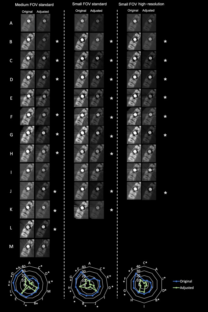
Background: The purpose of this study was to objectively assess dimensional alteration (blooming artefact) on dental implant using 13 cone-beam computed tomography (CBCT) devices adjusted to device-specific scanning protocols and to assess whether subjective adjustment of brightness and contrast (B&C) could alter its visualization.

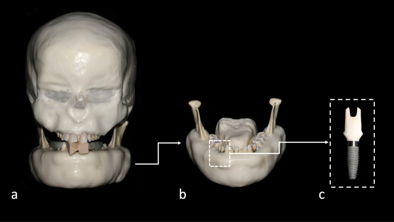
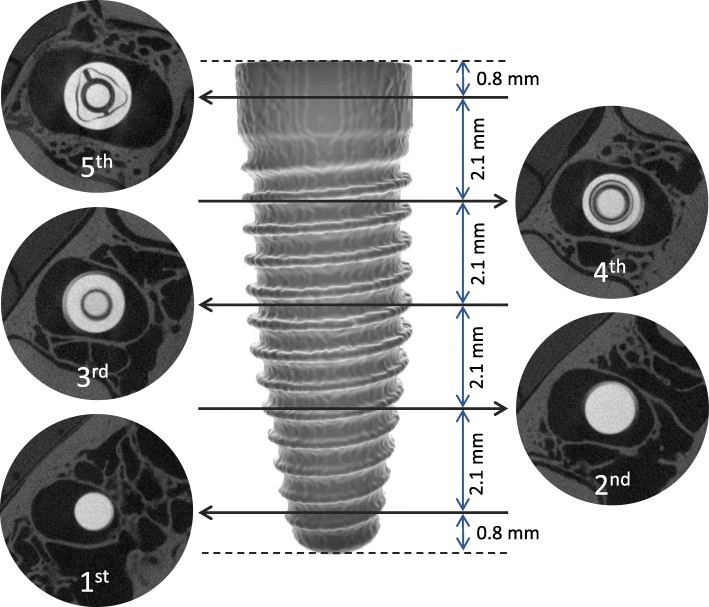
Methods: An anthropomorphic phantom containing a dental implant was scanned in 13 CBCT devices adjusted to three scanning protocols: medium-FOV standard resolution, small-FOV standard resolution, and small-FOV high resolution. The diameter of the implant was measured at five levels, averaged, and compared with those from a reference standard industrial CT image. B&C adjustments were performed and measurements were repeated. The intraclass correlation coefficient assessed the reliability of the measurements and general linear mixed models were applied for multiples comparisons at a 95% confidence interval.

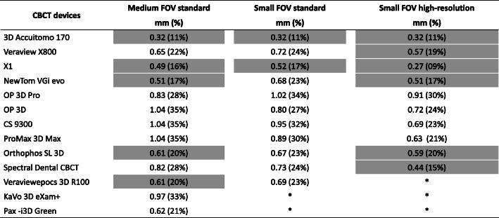
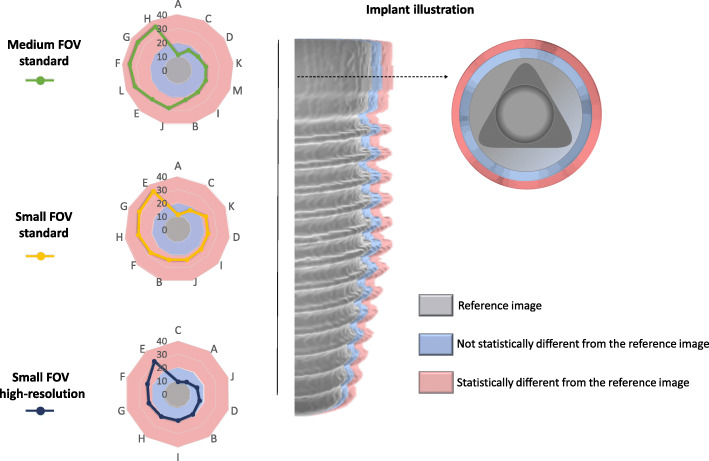
Results: Implant diameter obtained from small-FOV high-resolution protocols in most CBCT devices was not significantly different when compared to that from the reference (p > 0.05). For standard protocols, significant dimensional alteration of the implant ranging from 23 to 34% (0.67 to 1.02 mm) was observed in 9 CBCT devices for small-FOV scanning (p < 0.05), and in 8 CBCT devices for medium-FOV scanning, implant dimensional alteration ranged significantly from 21 to 35% (0.62 to 1.04 mm). After B&C adjustments, dimensional alteration was reduced for several of the CBCT devices tested (p < 0.05).

Conclusions: The visualization of the implant dimensional alteration differed between CBCT devices and scanning protocols with an increase in diameter ranging from 0.27 to 1.04 mm. For most CBCT devices, B&C adjustments allowed to reduce visualization of implant blooming.

Keywords: Artefacts; Cone-beam computed tomography; Dental implants.


Conflict of interest statement

Victor Aquino Wanderley, Karla de Faria Vasconcelos, Andre Ferreira Leite, Ruben Pauwels, Sohaib Shujaat, Reinhilde Jacobs, and Matheus L Oliveira declare that they have no competing interests.

Figures

Fig. 1
Fig. 1
Photographs of the imaging phantom. a Frontal view. b Frontal view of the mandible showing the tongue and implant of interest in place. c Magnified view of the studied implant
Fig. 2
Fig. 2
Illustration of the five implant levels and the corresponding axial slices from which the diameter was measured and averaged. The illustrated images were acquired in the industrial CT scanner (reference image)
Fig. 3
Fig. 3
Blooming artefact in millimetres and percentage increase of implant diameters compared with reference image for all studied CBCT devices and scanning protocols. The cells highlighted with light grey indicate that the implant measurements did not differ significantly from those of the reference image. Asterisks indicate the unavailable protocols
Fig. 4
Fig. 4
Left: Radar charts of the discrepancy (in percentage) of the diameter between the reference image and each CBCT device for the three scanning protocols. Right: An illustration of an implant (grey) with non-significant (blue) and significant (pink) expressions of the blooming artefact
Fig. 5
Fig. 5
Representative cropped axial slices of the original and B&C-adjusted images of all CBCT devices and scanning protocols. Asterisks indicate significant reduction of the negative impact of the blooming artefact after B&C adjustments. At the bottom of the figure, radar charts show the discrepancy (in percentage) of the implant diameter between the original and adjusted images of the CBCT devices for each device and scanning protocol

References

    1. Gaêta-Araujo H, Leite AF, Vasconcelos KF, Jacobs R. Two decades of research on CBCT imaging in DMFR - an appraisal of scientific evidence. Dentomaxillofac Radiol. 2021;50:20200367. 10.1259/dmfr.20200367. - PMC - PubMed
    1. Pauwels R, Stamatakis H, Bosmans H, Bogaerts R, Jacobs R, Horner K, Tsiklakis K, The SEDENTEXCT Project Consortium Quantification of metal artefacts on cone beam computed tomography images. Clin Oral Implants Res. 2013;24:94–99. doi: 10.1111/j.1600-0501.2011.02382.x. - DOI - PubMed
    1. Schulze R, Heil U, Gross D, Bruellmann DD, Dranischnikow E, Schwanecke U, et al. Artefacts in CBCT: a review. Dentomaxillofac Radiol. 2011;40(5):265–273. doi: 10.1259/dmfr/30642039. - DOI - PMC - PubMed
    1. Vasconcelos KF, Nicolielo LF, Nascimento MC, Haiter-Neto F, Bóscolo FN, Van Dessel J, et al. Artefact expression associated with several cone-beam computed tomographic machines when imaging root filled teeth. Int Endod J. 2015;48(10):994–1000. doi: 10.1111/iej.12395. - DOI - PubMed
    1. Codari M, de Faria VK, Ferreira Pinheiro Nicolielo L, Haiter Neto F, Jacobs R. Quantitative evaluation of metal artefacts using different CBCT devices, high-density materials and field of views. Clin Oral Implants Res. 2017;28(12):1509–1514. doi: 10.1111/clr.13019. - DOI - PubMed

Publication types

Substances

LinkOut - more resources