Clinical Trial
. 2021 Dec;41(12):3302-3313.
doi: 10.1177/0271678X211031035. Epub 2021 Jul 14.

Evaluation of [18F]-JNJ-64326067-AAA tau PET tracer in humans

Affiliations
Clinical Trial

Evaluation of [18F]-JNJ-64326067-AAA tau PET tracer in humans

Suzanne L Baker et al. J Cereb Blood Flow Metab. 2021 Dec.

Abstract

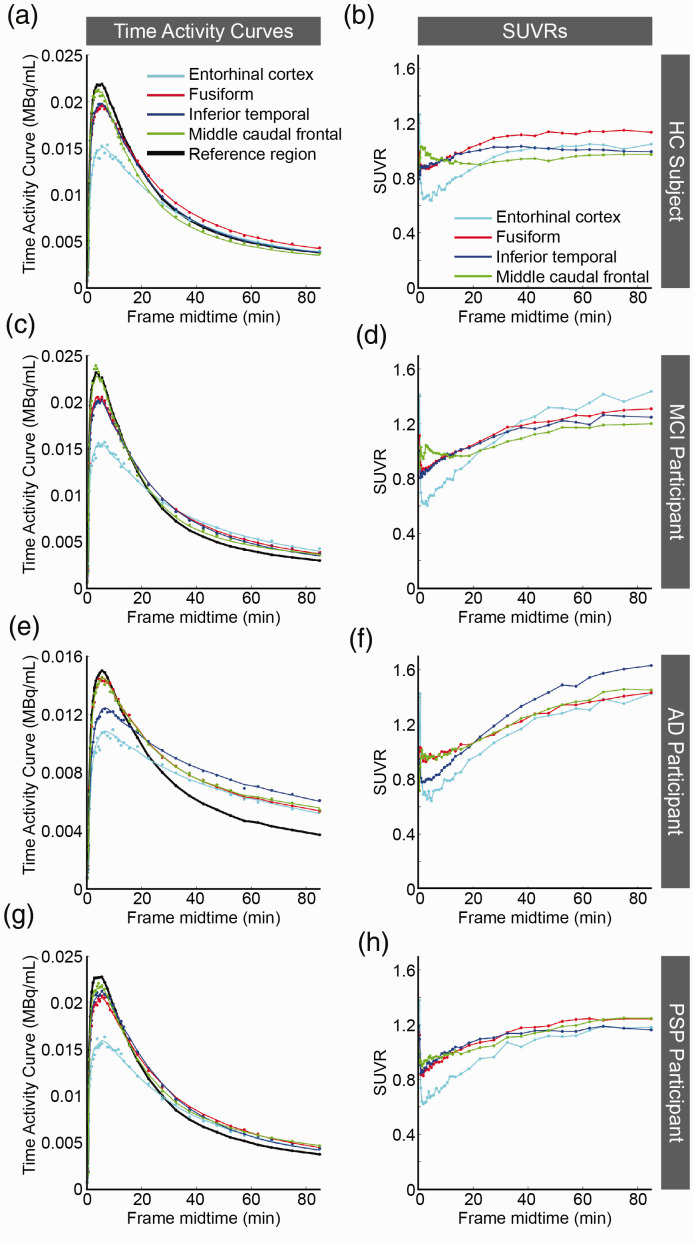
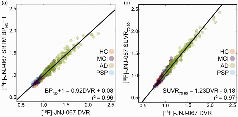
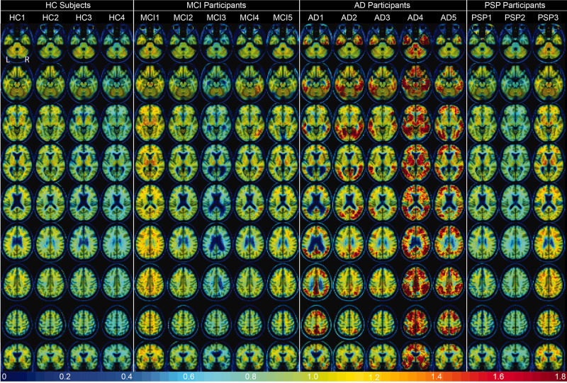
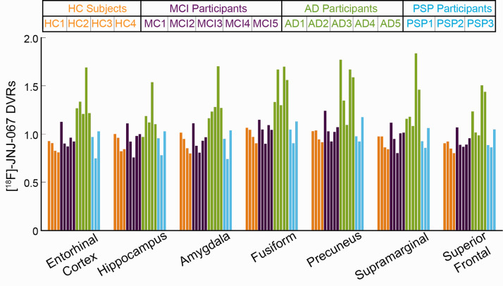
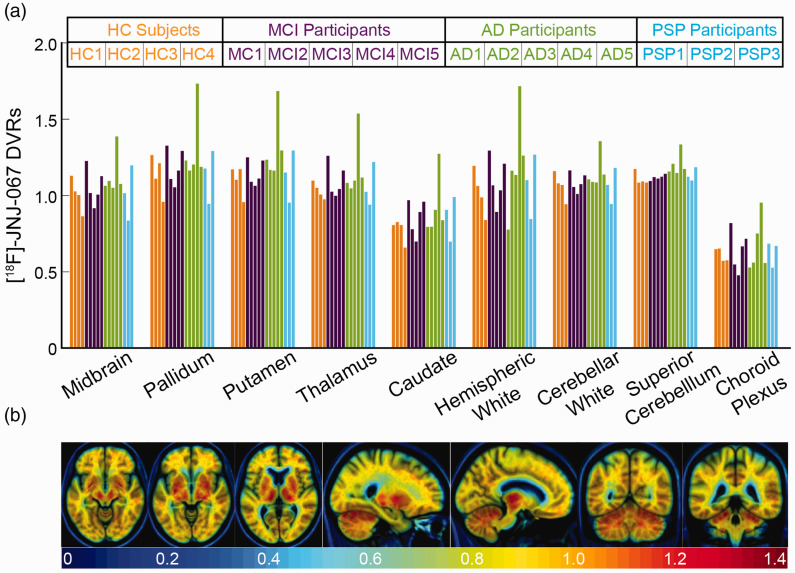
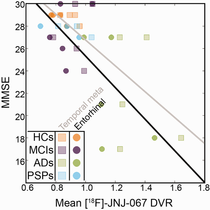
The [18F]-JNJ-64326067-AAA ([18F]-JNJ-067) tau tracer was evaluated in healthy older controls (HCs), mild cognitive impairment (MCI), Alzheimer's disease (AD), and progressive supranuclear palsy (PSP) participants. Seventeen subjects (4 HCs, 5 MCIs, 5 ADs, and 3 PSPs) received a [11C]-PIB amyloid PET scan, and a tau [18F]-JNJ-067 PET scan 0-90 minutes post-injection. Only MCIs and ADs were amyloid positive. The simplified reference tissue model, Logan graphical analysis distribution volume ratio, and SUVR were evaluated for quantification. The [18F]-JNJ-067 tau signal relative to the reference region continued to increase to 90 min, indicating the tracer had not reached steady state. There was no significant difference in any bilateral ROIs for MCIs or PSPs relative to HCs; AD participants showed elevated tracer relative to controls in most cortical ROIs (P < 0.05). Only AD participants showed elevated retention in the entorhinal cortex. There was off-target signal in the putamen, pallidum, thalamus, midbrain, superior cerebellar gray, and white matter. [18F]-JNJ-067 significantly correlated (p < 0.05) with Mini-Mental State Exam in entorhinal cortex and temporal meta regions. There is clear binding of [18F]-JNJ-067 in AD participants. Lack of binding in HCs, MCIs and PSPs suggests [18F]-JNJ-067 may not bind to low levels of AD-related tau or 4 R tau.

Keywords: Aging; Alzheimer’s disease; PET; [18F]-JNJ-067; tau.


Conflict of interest statement

Declaration of conflicting interests: The author(s) declared the following potential conflicts of interest with respect to the research, authorship, and/or publication of this article: Suzanne Baker consults for Genentech. Hartmuth Kolb and Mark Schmidt are employees of Janssen Research & Development. Maarten Timmers reports personal fees (current employment) from Janssen Research & Development, a Division of Janssen Pharmaceutica NV, Beerse, Belgium, and owns stock/stock options in the company. Gil Rabinovici receives research support from Avid, Eli Lily, GE Healthcare, and Life Molecular Imaging, serves as a consultant for Axon Neurosciences, Eisai, Merck, and GE, serves on the Data Safety Monitoring Board for Johnson & Johnson, and is associate editor of JAMA Neurology. William Jagust serves as a consultant for Genentech, Bioclinica, and Biogen and holds equity in Optoceutics.

Figures

Figure 1.
Figure 1.
Plots are time activity curves (TACs; a, c, e, g) and standardized uptake value ratios (SUVRs; b, d, f, h) for [18F]-JNJ-067. The data shown are from HC1 (a-b), MCI1 (c-d), AD1 (e-f), and PSP3 (g-h) (Table 1); all TACs and SUVRs are representative of their diagnostic groups except for MCI1 who had the greatest average mean cortical [18F]-JNJ-067 binding of all MCIs. The points represent mean PET values. The colored lines in a, c, e, and g are SRTM fits to the data. The lines in b, d, f and h just connect the individual points.
Figure 2.
Figure 2.
Correlation between DVR and SRTM BPND+1 (a) and DVR and SUVR70-90 (b) in 36 bilateral cortical ROIs across 17 subjects. Both correlations are significant (p<0.001) after Bonferroni correction.
Figure 3.
Figure 3.
[18F]-JNJ-067 DVR images (k2ref = 0.025 min−1; 35–90 min) warped to template space. Each column is a different subject following the order from Table 1, rows go through axial slices equally spaced for each subject ending with coronal showing entorhinal cortex.
Figure 4.
Figure 4.
Mean [18F]-JNJ-067 DVR values. Bars are ordered by HC (orange), MCI (purple), AD (green), and PSP (blue) subjects, and ordered within diagnosis group by increasing PIB SUVR index (as in Table 1).
Figure 5.
Figure 5.
(a) [18F]-JNJ-067 DVRs in regions that show off-target signal in other tau tracers. Bars are ordered by HC (orange), MCI (purple), AD (green), and PSP (blue) subjects, and ordered within diagnosis group by increasing PIB SUVR index (as in Table 1). (b) Mean of 4 HC subjects in template space showing regions of potential off-target signal.
Figure 6.
Figure 6.
[18F]-JNJ-067 entorhinal cortex DVR vs MMSE (circles; r = −0.52; p < 0.05) and temporal meta ROI vs MMSE (squares, r= −0.56; p < 0.05).

References

    1. Braak H, Braak E. Neuropathological stageing of Alzheimer-related changes. Acta Neuropathol 1991; 82: 239–259. - PubMed
    1. Chien DT, Bahri S, Szardenings AK, et al.. Early clinical PET imaging results with the novel PHF-tau radioligand [F-18]-T807. J Alzheimers Dis 2013; 34: 457–468. - PubMed
    1. Kuwabara H, Comley RA, Borroni E, et al.. Evaluation of (18)F-RO-948 PET for quantitative assessment of tau accumulation in the human brain. J Nucl Med 2018; 59: 1877–1884. - PMC - PubMed
    1. Wong DF, Comley RA, Kuwabara H, et al.. Characterization of 3 novel tau radiopharmaceuticals, (11)C-RO-963, (11)C-RO-643, and (18)F-RO-948, in healthy controls and in Alzheimer subjects. J Nucl Med 2018; 59: 1869–1876. - PMC - PubMed
    1. Hostetler ED, Walji AM, Zeng Z, et al.. Preclinical characterization of 18F-MK-6240, a promising PET tracer for in vivo quantification of human neurofibrillary tangles. J Nucl Med 2016; 57: 1599–1606. - PubMed

Publication types