. 2022 Apr;28(4):522-531.
doi: 10.1177/13524585211028831. Epub 2021 Jul 14.

Optical coherence tomography angiography indicates subclinical retinal disease in neuromyelitis optica spectrum disorders

Affiliations

Optical coherence tomography angiography indicates subclinical retinal disease in neuromyelitis optica spectrum disorders

Lilian Aly et al. Mult Scler. 2022 Apr.

Abstract

Background: Neuromyelitis optica spectrum disorders (NMOSD) are neuroinflammatory diseases of the central nervous system. Patients suffer from recurring relapses and it is unclear whether relapse-independent disease activity occurs and whether this is of clinical relevance.

Objective: To detect disease-specific alterations of the retinal vasculature that reflect disease activity during NMOSD.

Methods: Cross-sectional analysis of 16 patients with NMOSD, 21 patients with relapsing-remitting multiple sclerosis, and 21 healthy controls using retinal optical coherence tomography (OCT), optical coherence tomography angiography (OCT-A), measurement of glial fibrillary acidic protein (GFAP) serum levels, and assessment of visual acuity.

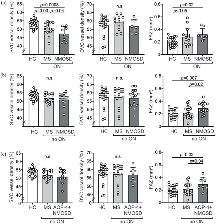
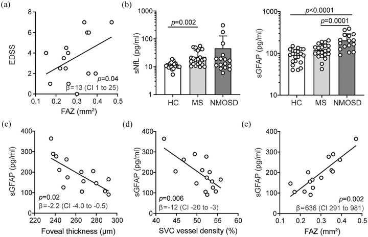
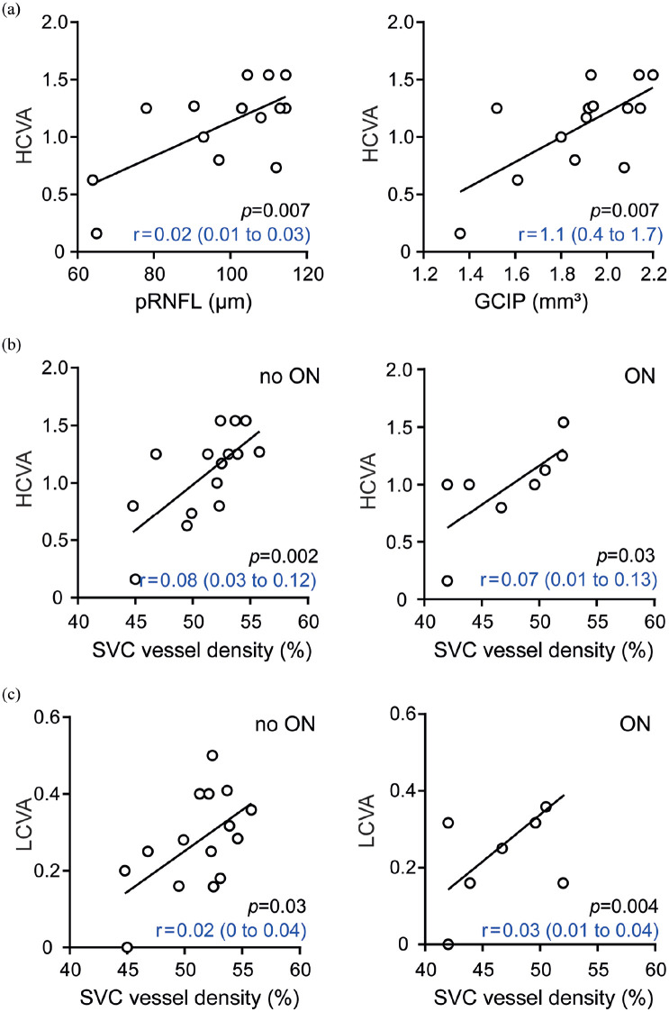
Results: Patients with NMOSD but not multiple sclerosis revealed lower foveal thickness (FT) (p = 0.02) measures and an increase of the foveal avascular zone (FAZ) (p = 0.02) compared to healthy controls independent to optic neuritis. Reduced FT (p = 0.01), enlarged FAZ areas (p = 0.0001), and vessel loss of the superficial vascular complex (p = 0.01) were linked to higher serum GFAP levels and superficial vessel loss was associated with worse visual performance in patients with NMOSD irrespective of optic neuritis.

Conclusion: Subclinical parafoveal retinal vessel loss might occur during NMOSD and might be linked to astrocyte damage and poor visual performance. OCT-A may be a tool to study subclinical disease activity during NMOSD.

Keywords: Neuromyelitis optica spectrum disorders; astrocytes; biomarker; disease activity; optical coherence tomography angiography.


Conflict of interest statement

Declaration of Conflicting Interests: The author(s) declared the following potential conflicts of interest with respect to the research, authorship, and/or publication of this article: L.A. received travel support and a poster grant from Novartis. A.B. received speaker and consulting honoraria from Alexion, Biogen, Bayer Healthcare, Celgene, Merck, Novartis Pharma, and Roche, all outside the submitted work. B.H. has served on scientific advisory boards for Novartis, he has served as DMSC member for AllergyCare, Polpharma, and TG therapeutics, he or his institution have received speaker honoraria from Desitin, his institution received research grants from Regeneron for MS research, he holds part of two patents, one for the detection of antibodies against KIR4.1 in a subpopulation of patients with MS and one for genetic determinants of neutralizing antibodies to interferon, all conflicts are not relevant to the topic of the study. M.M. reports research funding from Novartis, Bayer, and Roche, he received speaker honoraria from Allergan, Bayer, Novartis, and Heidelberg Engineering, all conflicts are not relevant to the topic of the study. B.K. received travel support and a research grant from Novartis (Oppenheim research award). E.-M.S., N.F., I.W., M.M., C.H., and T.K. report no disclosures.

Figures

Figure 1.
Figure 1.
Results of optical coherence tomography angiography analysis. (a) Parafoveal vessel densities of the superficial (SVC) and deep vascular complex (DVC) and size of the foveal avascular zone (FAZ) in healthy individuals (HC, n = 21) and eyes with former optic neuritis (ON) of patients with relapsing-remitting MS (MS, n = 14 patients) and neuromyelitis optica spectrum disorders (NMOSD, n = 8 patients); one eye of one NMOSD patient was excluded due to poor OCT quality. (b) Parafoveal vessel densities of the SVC and DVC and size of the FAZ in HC (n = 21), MS (n = 18 patients), and NMOSD (n = 15 patients) in eyes without a history of optic neuritis (no ON); one MS patient underwent unilateral enucleation in the past. (c) Parafoveal vessel densities of the SVC and DVC and size of the FAZ in HC (n = 21), MS (n = 18 patients), and NMOSD with antibodies against aquaporin-4 (AQP-4 + NMOSD) (n = 9 patients) in eyes without a history of optic neuritis (no ON). (a–c) Mean ± standard deviation (SD); symbols depict single patient values; one-way ANOVA; n.s.: not significant; the statistical significance threshold was p<0.05.
Figure 2.
Figure 2.
Retinal architecture, vasculature, and surrogate markers of disease activity in neuromyelitis optica spectrum disorders. (a) Association of the Expanded Disability Status Scale (EDSS) and the foveal avascular zone (FAZ) areas during neuromyelitis optica spectrum disorders (NMOSD) (n = 15); β regression estimate and 95% confidence interval (CI); symbols depict single patients; multiple linear regression model corrected for age and sex. (b) Serum levels of neurofilament light chain (sNfL) and glial fibrillary acidic protein (sGFAP) in healthy individuals (HC, n = 21), patients with relapsing-remitting MS (MS, n = 21) and NMOSD (n = 15); mean ± standard deviation; symbols depict single patients; Kruskal–Wallis test (sNfL), one-way ANOVA (GFAP). (c–e) Association of sGFAP levels and foveal thickness (FT) (c), vessel densities of the superficial vascular complex (SVC) (d), and the foveal avascular zone (FAZ) areas (e) in eyes without former optic neuritis in patients with NMOSD (n = 15); β regression estimates and 95% CI; symbols depict single patients; multiple linear regression models corrected for age, sex, and EDSS.
Figure 3.
Figure 3.
Retinal architecture, vasculature, and visual function in neuromyelitis optica spectrum disorders. (a) Association of high-contrast visual acuity (HCVA) and thickness of the peripapillary retinal nerve fiber layer (pRNFL, left) or volumes of the common ganglion cell and inner plexiform layer (GCIP, right) in eyes without former optic neuritis (ON) in patients with neuromyelitis optica spectrum disorders (NMOSD) (n = 14). (b) Association of HCVA and vessel densities of the superficial vascular complex (SVC) in eyes of NMOSD patients without (n = 15, left) or with a history of optic neuritis (n = 8, right). (c) Association of low-contrast visual acuity (LCVA) and vessel densities of SVC in eyes of NMOSD patients without (n = 15, left) or with a history of optic neuritis (n = 8, right). (a–c) β regression estimates and 95% CI; symbols depict single patients; multiple linear regression model corrected for age and sex.

References

    1. Jarius S, Paul F, Weinshenker BG, et al.. Neuromyelitis optica. Nat Rev Dis Primers 2020; 6: 85. - PubMed
    1. Oertel FC, Kuchling J, Zimmermann H, et al.. Microstructural visual system changes in AQP4-antibody-seropositive NMOSD. Neurol Neuroimmunol Neuroinflamm 2017; 4(3): e334. - PMC - PubMed
    1. Motamedi S, Oertel FC, Yadav SK, et al.. Altered fovea in AQP4-IgG-seropositive neuromyelitis optica spectrum disorders. Neurol Neuroimmunol Neuroinflamm 2020; 7(5): e805. - PMC - PubMed
    1. Roca-Fernandez A, Oertel FC, Yeo T, et al.. Foveal changes in aquaporin-4 antibody seropositive neuromyelitis optica spectrum disorder are independent of optic neuritis and not overtly progressive. Eur J Neurol 2021; 28(7): 2280–2293. - PubMed
    1. Filippatou AG, Vasileiou ES, He Y, et al.. Evidence of subclinical quantitative retinal layer abnormalities in AQP4-IgG seropositive NMOSD. Mult Scler. Epub ahead of print 14 December 2020. DOI: 10.1177/1352458520977771. - DOI - PMC - PubMed

Publication types