

SARS-CoV-2 B.1.617 Indian variants: Are electrostatic potential changes responsible for a higher transmission rate?
- PMID: 34260088
- PMCID: PMC8426736
- DOI: 10.1002/jmv.27210
SARS-CoV-2 B.1.617 Indian variants: Are electrostatic potential changes responsible for a higher transmission rate?
Abstract
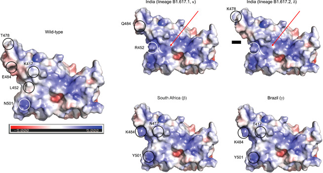
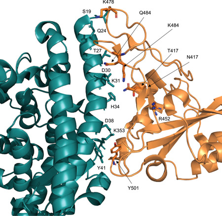
Lineage B.1.617+, also known as G/452R.V3 and now denoted by WHO with the Greek letters δ and κ, is a recently described SARS-CoV-2 variant under investigation first identified in October 2020 in India. As of May 2021, three sublineages labeled as B.1.617.1 (κ), B.1.617.2 (δ), and B.1.617.3 have been already identified, and their potential impact on the current pandemic is being studied. This variant has 13 amino acid changes, three in its spike protein, which are currently of particular concern: E484Q, L452R, and P681R. Here, we report a major effect of the mutations characterizing this lineage, represented by a marked alteration of the surface electrostatic potential (EP) of the receptor-binding domain (RBD) of the spike protein. Enhanced RBD-EP is particularly noticeable in the B.1.617.2 (δ) sublineage, which shows multiple replacements of neutral or negatively charged amino acids with positively charged amino acids. We here hypothesize that this EP change can favor the interaction between the B.1.617+ RBD and the negatively charged ACE2, thus conferring a potential increase in the virus transmission.
Keywords: B.1.617 δ and κ variants; SARS-CoV-2; electrostatics potential changes.
© 2021 The Authors. Journal of Medical Virology Published by Wiley Periodicals LLC.
Figures
References
-
- Wang C, Horby PW, Hayden FG, Gao GF. A novel coronavirus outbreak of global health concern. Lancet. 2020;395:P470‐P473. https://www.thelancet.com/journals/lancet/article/PIIS0140-6736(20)30185... - PMC - PubMed
MeSH terms
Substances
LinkOut - more resources
Full Text Sources
Medical
Research Materials
Miscellaneous
