. 2021 Jul 28;69(29):8120-8129.
doi: 10.1021/acs.jafc.1c01876. Epub 2021 Jul 14.

Structure and Zeatin Binding of the Peach Allergen Pru p 1

Affiliations

Structure and Zeatin Binding of the Peach Allergen Pru p 1

Reiner Eidelpes et al. J Agric Food Chem. .

Abstract

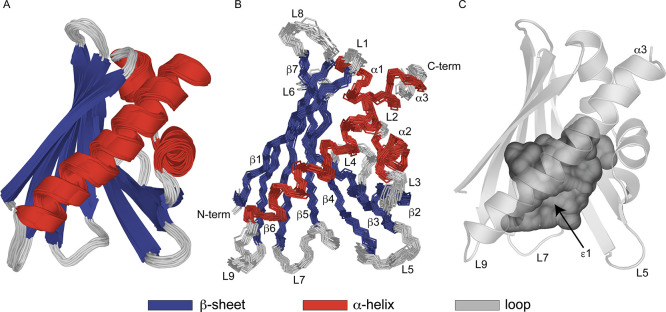
Peach (Prunus persica) is among the fruits most frequently reported to cause food allergies. Allergic reactions commonly result from previous sensitization to the birch pollen allergen Bet v 1, followed by immunological cross-reactivity of IgE antibodies to structurally related proteins in peach. In this study, we present the three-dimensional NMR solution structure of the cross-reactive peach allergen Pru p 1 (isoform Pru p 1.0101). This 17.5 kDa protein adopts the canonical Bet v 1 fold, composed of a seven-stranded β-sheet and three α-helices enclosing an internal cavity. In Pru p 1, the inner surface of the cavity contains an array of hydroxyl-bearing amino acids surrounded by a hydrophobic patch, constituting a docking site for amphiphilic molecules. NMR-guided docking of the cytokinin molecule zeatin to the internal cavity of Pru p 1 provides a structure-based rationale for the effect that zeatin binding has on the protein's RNase activity.

Keywords: NMR structure; allergen; cytokinin; ribonuclease.


Conflict of interest statement

The authors declare no competing financial interest.

Figures

Figure 1
Figure 1
NMR solution structure of the peach allergen Pru p 1.0101 (pdb accession code 6Z98). (A) Ribbon representation of the ensemble of the 20 lowest energy structures. Secondary structure elements β1 (Val2-Ser11), β2 (Ile38-Glu45), β3 (Gly51-Phe58), β4 (Gly65-Asp75), β5 (His79-Ile86), β6 (Leu95-Ala106), β7 (Ser112-Thr122), α1 (Pro15-Val23), α2 (Ala26-Ala34), and α3 (Glu130-His154) are colored in blue and red. (B) Backbone overlay of the 20 lowest energy structures. Secondary structure elements, loops (L1–L9) and N- and C-termini are labeled. (C) Internal cavity of Pru p 1.0101, which is accessible via the entrance ε1 formed by the N-terminal end of helix α3 and loops L5, L7, and L9. Figures were prepared using the program PYMOL.
Figure 2
Figure 2
Internal cavity of Pru p 1.0101. (A) Strands β1 and β4−β7 contain nine amino acid residues (shown in orange), whose side chain hydroxyl groups are oriented toward the inner surface of the cavity, which are flanked by hydrophobic residues (yellow). The single histidine residue, His69, is shown in green. (B) Opposite face of the cavity is formed by hydrophobic residues in helices α1−α3, strands β2−β3 and loops L2 and L3. For illustrative purposes, the backbone of helix α3, which covers the internal cavity, is transparent in both panels.
Figure 3
Figure 3
Binding of trans-zeatin to the peach allergen Pru p 1.0101. (A) Sections from backbone amide 1H–15N HSQC spectra of Pru p 1 (0.2 mM) in the presence of variable amounts (up to 16-fold excess) of zeatin, recorded at 500 MHz. Data of four representative amino acid residues (Leu24, Lys68, Tyr81, and Ser141) are shown. (B) Zeatin binding curves for four representative amino acid residues. Kd values for zeatin binding to Pru p 1.0101 were derived from 15N chemical shift changes, Δδ(15N), of these residues upon ligand binding by nonlinear least squares fitting of the binding curves as described. Additional experimental data for residues with Δδ(15N) exceeding 0.15 ppm are provided in Supporting Information Figure S2.
Figure 4
Figure 4
Interaction site of trans-zeatin. (A) Protein backbone of Pru p 1.0101 as viewed from the entrance to the internal cavity, ε1. Backbone amides with significant CSPs or increased 15N RD rates (Rex) upon zeatin binding are represented as purple and cyan spheres, respectively. Side chain 1Hδ and 1Hε nuclei of phenylalanines and tyrosines with CSP are displayed as green spheres. (B) Schematic and (C) detailed structural representation of the molecular docking model. Side chains of amino acids surrounding zeatin (distance ≤ 3.5 Å) are displayed in orange (tyrosine and histidine) and yellow (hydrophobic residues), while zeatin is shown in green. Red dashes represent hydrogen bonds from the imidazole side chain of His69 to N1 and from the side chain hydroxyl group of Tyr81 to N3 of the adenine moiety of zeatin. Blue dashes indicate the π-stacking interaction between the aromatic side chain of Phe22 and the purine ring system of zeatin. For illustrative purposes, in panel (C), helices α1 and α2 and Ala26 in loop L2 are transparent.
Figure 5
Figure 5
Structure of the β2–L4−β3 segment in Pru p 1.0101 encompassing the glycine-rich region, Gly46-Gly51. (A) Lowest energy structure of the glycine rich loop. Hydrogen bonds are indicated as red dashed lines. (B) Superposition of the β2–L4−β3 segment in Pru p 1 (structural bundle, gray) with Bet v 1 (yellow) in complex with the antibody fragment Fab of BV16 (pale green, pdb entry code 1FSK). The side chains of Pru p 1 residues between Glu45 and Gly51 are shown as sticks.

References

    1. Burney P. G. J.; Potts J.; Kummeling I.; Mills E. N. C.; Clausen M.; Dubakiene R.; Barreales L.; Fernandez-Perez C.; Fernandez-Rivas M.; Le T.-M.; Knulst A. C.; Kowalski M. L.; Lidholm J.; Ballmer-Weber B. K.; Braun-Fahlander C.; Mustakov T.; Kralimarkova T.; Popov T.; Sakellariou A.; Papadopoulos N. G.; Versteeg S. A.; Zuidmeer L.; Akkerdaas J. H.; Hoffmann-Sommergruber K.; van Ree R. The prevalence and distribution of food sensitization in European adults. Allergy 2014, 69, 365–371. 10.1111/all.12341. - DOI - PubMed
    1. Ma S.; Yin J.; Jiang N. Component-resolved diagnosis of peach allergy and its relationship with prevalent allergenic pollens in China. J. Allergy Clin. Immunol. 2013, 132, 764–767. 10.1016/j.jaci.2013.04.017. - DOI - PubMed
    1. Geroldinger-Simic M.; Zelniker T.; Aberer W.; Ebner C.; Egger C.; Greiderer A.; Prem N.; Lidholm J.; Ballmer-Weber B. K.; Vieths S.; Bohle B. Birch pollen-related food allergy: clinical aspects and the role of allergen-specific IgE and IgG4 antibodies. J. Allergy Clin. Immunol. 2011, 127, 616–622. 10.1016/j.jaci.2010.10.027. - DOI - PubMed
    1. Ma S.; Sicherer S. H.; Nowak-Wegrzyn A. A survey on the management of pollen-food allergy syndrome in allergy practices. J. Allergy Clin. Immunol. 2003, 112, 784–788. 10.1016/s0091-6749(03)02008-6. - DOI - PubMed
    1. Wisniewski M.; Bassett C.; Arora R. Distribution and partial characterization of seasonally expressed proteins in different aged shoots and roots of ’Loring’ peach (Prunus persica). Tree Physiol. 2004, 24, 339–345. 10.1093/treephys/24.3.339. - DOI - PubMed