Case Reports
. 2021 Jul 16;100(28):e26445.
doi: 10.1097/MD.0000000000026445.

Similar appearance of different multifocal carpal bone destructing disease entities in 3 patients: A case report

Affiliations
Case Reports

Similar appearance of different multifocal carpal bone destructing disease entities in 3 patients: A case report

Jun-Ku Lee et al. Medicine (Baltimore). .

Abstract

Rationale: Several diseases feature tumors, or tumor-mimicking lesions, that further invade the bone and surrounding joints of the wrist region. Here, we describe 3 rare cases of multiple destructed carpal bones and adjacent joints in different disease entities confirmed via pathologic diagnosis.

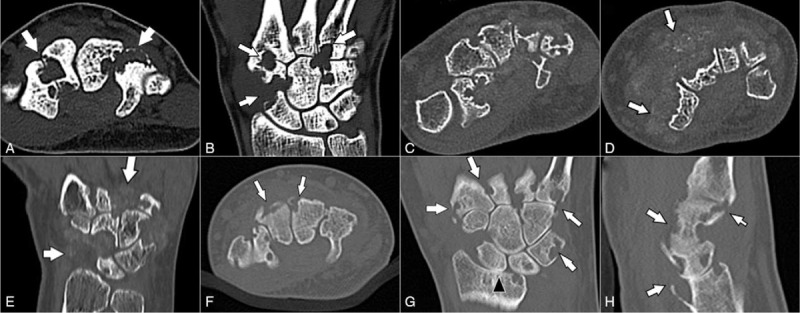
Patient concerns: All 3 cases were examined between January 2016 and December 2019. Three patients presented with similar clinical manifestations and radiographic features, with multiple osteolytic lesions in the carpal bones and metacarpal bone base.

Diagnoses: The 3 cases were diagnosed as diffuse type tenosynovial giant cell tumor, calcifying aponeurotic fibroma, and rheumatoid arthritis.

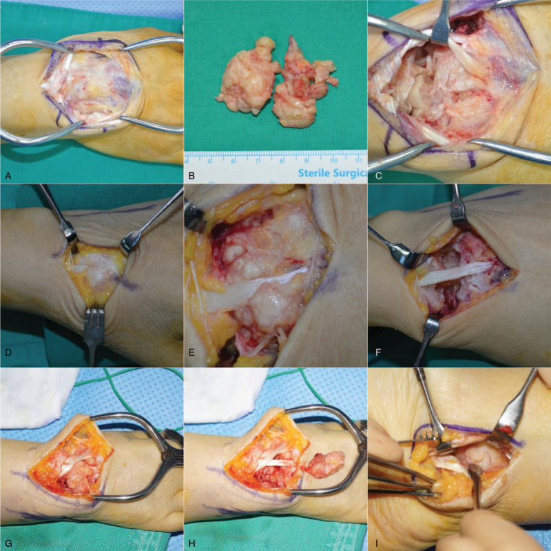
Interventions: Separate, experienced radiologist and pathologist took part in the interpretation and compartmentalization of radiographs and pathological findings, respectively. Even magnetic resonance imaging could not achieve a diagnosis; surgical excision was therefore required, with subsequent pathological assessment for treatment and final diagnosis.

Outcomes: functional outcomes also differed among patients, poorest in rheumatoid arthritis patient.

Lessons: We report 3 rare disease entities, presenting with multifocal osteolytic lesions in the wrist. They all presented with similar clinical manifestations, and the final diagnoses were made via pathological evaluation. Compared with tenosynovial giant cell tumor and calcifying aponeurotic fibroma, rheumatoid arthritis had the poorest outcome.


Conflict of interest statement

The authors report no conflicts of interest.

Figures

Figure 1
Figure 1
Plain x-ray, anteroposterior (AP), and lateral views of the 3 patients. Plain radiographs of Case 1 (A and B) reveal multiple well-defined carpal and metacarpal base erosions (arrows). In Case 2, the AP (C) and lateral views (D) show multiple erosions of the carpal bones and metacarpal bases, with surrounding, ill-defined, lobulated soft tissue density (arrows). Case 3 (E and F) also reveals multiple erosions of the radius, carpal bones, and metacarpal bases (arrows), with narrowing of the radiocarpal joint space.
Figure 2
Figure 2
Computed tomography images of the 3 patients. In Case 1, the axial (A) and coronal (B) images reveal multiple erosions of the carpal bones and metacarpal bases (arrows). The axial (C and D) and coronal (E) images of Case 2 showed multiple erosions of the carpal bones and metacarpal bases; surrounding, ill-defined, lobulated, soft tissue mass-like lesions show stippled calcification (arrows). The axial (F), coronal (G), and sagittal (H) images of Case 3 demonstrate multiple erosions of the radius, carpal bones, and metacarpal bases (arrows), with narrowing of the radiocarpal joint space (arrowhead).
Figure 3
Figure 3
Magnetic resonance images of the 3 patients. In Case 1, multiple nodular, synovial, proliferative lesions, causing erosion of the carpal bones and metacarpal bases, revealed a low signal intensity on coronal T1-weighted imaging (A), and a heterogeneous high signal intensity on coronal fat-suppressed T2-weighted imaging (B) (arrows). In Case 2, multiple nodular, synovial, proliferative lesions, causing erosion of the carpal bones and metacarpal bases, showed a low signal intensity on coronal T1-weighted imaging (C), heterogeneous high signal intensity on coronal fat-suppressed T2-weighted imaging (D), and heterogeneous enhancement (E) (arrows). In Case 3, diffuse, synovial, proliferative lesions, causing erosion of the radius, carpal bones, and metacarpal bases demonstrated a heterogeneous high signal intensity on coronal intermediate (modified proton density-weighted) imaging (F) and fat-suppressed T2-weighted imaging (G), and heterogeneous enhancement (H) (arrows). The radiocarpal joint shows cartilage loss and joint space narrowing, with distal radioulnar compartment effusion. Diffuse bone marrow edema is seen in the distal radius and ulna, carpal bones, and metacarpal bases.
Figure 4
Figure 4
Intraoperative clinical photos of the 3 patients. In the first patient, the tumor was buried in the carpal bone and CMC joint; the articular capsule and boundary under the extensor tendon were unclear (A). Marginal excision was performed to remove all potential masses (B and C). In the second patient, the mass was found in the subcutaneous layer, enveloping the extensor tendons (D). The calcified mass invading the surrounding retinaculum and joint capsule was not distinguished (E), and was detached from the bone and adjacent joints (F). In the third patient, solid, mass-like synovium invaded the retinaculum, joint capsule, underlying bone, and joints with intact tendon (G), and was subsequently removed (H). A separated volar approach was used to remove the volar aspect lesion (I).
Figure 5
Figure 5
Pathological findings of the 3 patients. Case 1: giant cell tumor of the tendon sheath (A and B); Case 2: calcifying aponeurotic fibroma (C and D); Case 3; rheumatoid arthritis (E, F, and G). (A) Moderately cellular tumor showing abundant mononuclear cells and evenly scattered, multinucleated, osteoclast-like giant cells (arrows) within hyalinized fibrous stroma (HE ×40). (B) The mononuclear component of the tumor contains small to medium sized cells, with ovoid nuclei and a pale cytoplasm. Osteoclast-like giant cells (arrows) are occasionally admixed (HE ×200). (C) Zone of hyalinized chondroid matrix and chondrocyte-like cells with calcified nodules (HE ×100). (D) Cellular area showing plump/epithelioid to spindle cells, osteoclast-like giant cells (arrows), and spotty calcification (arrowheads) (HE ×200). (E) Proliferative synovitis with villous hypertrophy, and dense inflammatory cell infiltration with lymphoid follicles (arrows) (HE ×40). (F) Presence of fibrinoid necrosis (arrowheads) (HE ×40). (G) Inflammatory infiltrates are primarily composed of lymphocytes (upper left) and plasma cells (lower right) (HE ×200).

References

    1. Garcia-Elias M, Lluch AL. Cohen MS, Green DP, Hotchkiss RN, Kozin SH, Pederson WC, Wolfe SW. Wrist instabilities, misalignments, and dislocations. Green's Operative Hand Surgery.. Philadelphia, PA: Elsevier; 2017. 418–78.
    1. Zhao X, Qi C, Chen J, et al. . Synovial hemangioma of the wrist with cystic invasion of trapezoid and capitate bones. J Hand Surg Am 2020;45:161.e1–e6. - PubMed
    1. Lee SM, Ha DH, Kang H, et al. . Intraarticular calcifying aponeurotic fibroma of the wrist: mimicking gout or calcium pyrophosphate dihydrate deposition disease. Skeletal Radiol 2018;47:729–34. - PubMed
    1. Kim OH, Kim YM. Calcifying aponeurotic fibroma: case report with radiographic and MR features. Korean J Radiol 2014;15:134–9. - PMC - PubMed
    1. Athanasian EA. Cohen MS, Green DP, Hotchkiss RN, Kozin SH, Pederson WC, Wolfe SW. Bone and soft tissue tumors. Green's Operative Hand Surgery. Philadelphia, PA: Elsevier; 2017. 1987–2035.

Publication types

MeSH terms

Supplementary concepts