. 2021 Jul 13;36(2):109383.
doi: 10.1016/j.celrep.2021.109383.

Protein phosphatase 1 acts as a RIF1 effector to suppress DSB resection prior to Shieldin action

Affiliations

Protein phosphatase 1 acts as a RIF1 effector to suppress DSB resection prior to Shieldin action

Shin-Ya Isobe et al. Cell Rep. .

Abstract

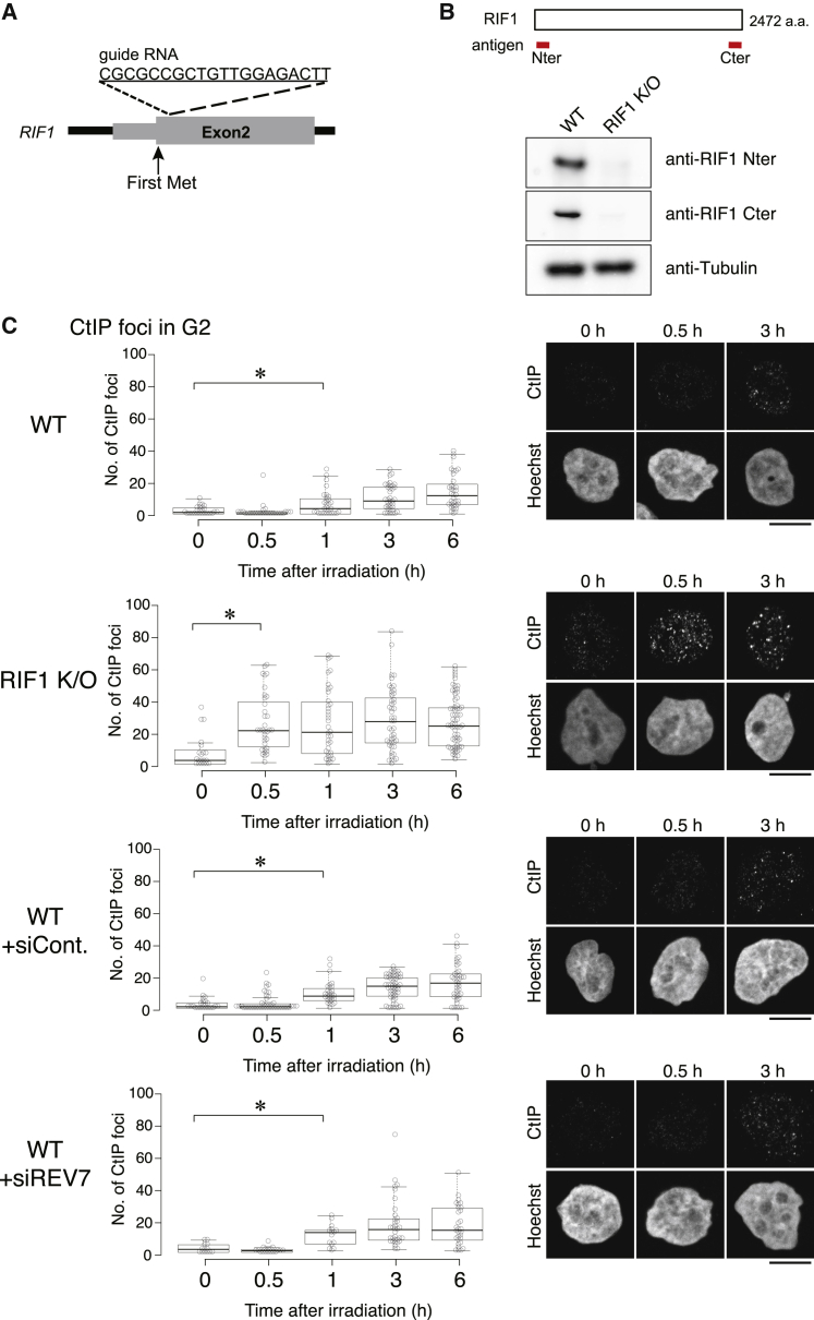
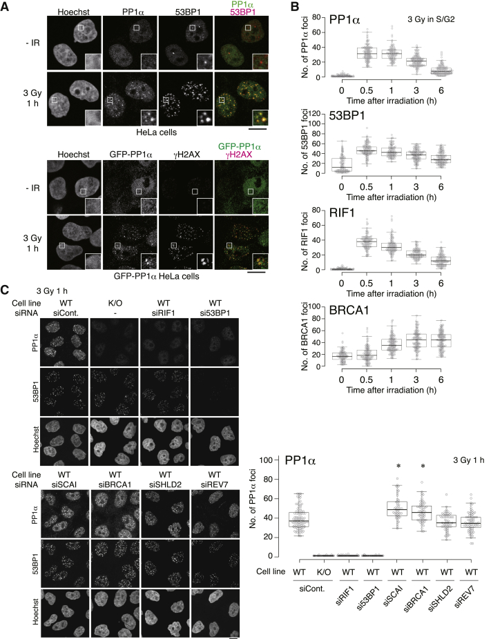
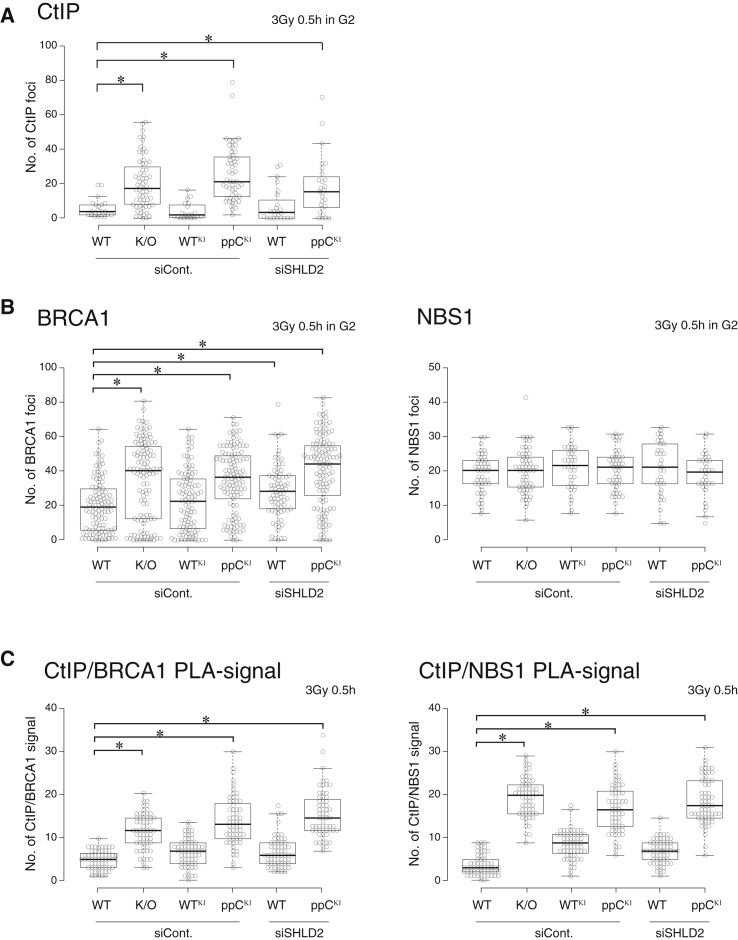
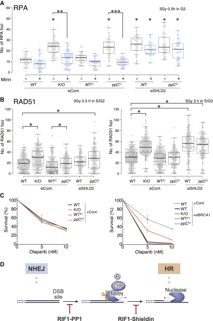
DNA double-strand breaks (DSBs) are repaired mainly by non-homologous end joining (NHEJ) or homologous recombination (HR). RIF1 negatively regulates resection through the effector Shieldin, which associates with a short 3' single-stranded DNA (ssDNA) overhang by the MRN (MRE11-RAD50-NBS1) complex, to prevent further resection and HR repair. In this study, we show that RIF1, but not Shieldin, inhibits the accumulation of CtIP at DSB sites immediately after damage, suggesting that RIF1 has another effector besides Shieldin. We find that protein phosphatase 1 (PP1), a known RIF1 effector in replication, localizes at damage sites dependent on RIF1, where it suppresses downstream CtIP accumulation and limits the resection by the MRN complex. PP1 therefore acts as a RIF1 effector distinct from Shieldin. Furthermore, PP1 deficiency in the context of Shieldin depletion elevates HR immediately after irradiation. We conclude that PP1 inhibits resection before the action of Shieldin to prevent precocious HR in the early phase of the damage response.

Keywords: CtIP; HR; MRN; NHEJ; Olaparib; PP1; Pathway choice; RIF1; Resection; Shieldin.


Conflict of interest statement

Declaration of interests The authors declare no competing interests.

Figures

None
Graphical abstract
Figure 1
Figure 1
RIF1, but not Shieldin, suppresses CtIP IRIF (A) Schematic diagram of RIF1 gene construct on the genome, with guide RNA sequence for disruption by CRISPR-Cas9. (B) Western blot of whole-cell extracts prepared from wild-type (WT) cells and RIF1 knockout (KO) cells, probed with indicated antibodies. Tubulin is a loading control. (C) CtIP IRIF in the G2 phase. Indicated cell lines were treated with/without indicated siRNA for 48 h and then irradiated. After irradiation (3 Gy), cells were fixed at the indicated time (with addition of 5-ethynyl-2′-deoxyuridine [EdU] 10 min beforehand) and immunostained with an anti-CtIP antibody, along with Hoechst staining and EdU Click-iT visualization. Cells in the G2 phase were assigned based on Hoechst intensity and lack of EdU signal. The numbers of CtIP foci in the G2 phase are shown as bee swarm plots (left). p values were calculated using a Mann-Whitney U test (p < 0.001). Representative images are shown for cells 0, 0.5, and 3 h post-irradiation (right). Scale bars, 10 μm. See also Figure S1.
Figure 2
Figure 2
PP1 IRIF depends on RIF1 (A) PP1α IRIF. HeLa cells (upper) or HeLa cells stably expressing GFP-PP1α (lower) were irradiated (3 Gy) and then 1 h later fixed for immunostaining with anti-PP1α, anti-53BP1, and anti-γH2AX antibodies. GFP-PP1α was detected as GFP signal. Scale bars, 10 μm. (B) Time course analysis of IRIF for PP1α, 53BP1, RIF1, and BRCA1 in S/G2 cells. Cells were irradiated (3 Gy) and incubated for the indicated periods prior to fixation for immunofluorescence. Cells were immunostained with antibodies against indicated proteins and cyclin A, along with Hoechst staining. Cells in the S/G2 phase were assigned based on cyclin A intensity. Numbers of foci are shown as a bee swarm plot. (C) Dependency of PP1 IRIF on RIF1 and 53BP1. WT or RIF1 KO cells were treated with indicated siRNA for 48 h and irradiated (3 Gy) and then 1 h later fixed for immunostaining with anti-PP1α and anti-53BP1 antibodies. The numbers of PP1α foci are shown as bee swarm plots (p < 0.001 for WT siCont). Scale bar, 10 μm. See also Figure S2.
Figure 3
Figure 3
PP1 IRIF depends on the direct binding to RIF1 (A) Schematic structure of RIF1 indicating its three PP1 binding motifs and the mutants created. (B) Testing PP1 binding for the mutants created. T-REx 293 cells were transfected with expression plasmids for the indicated constructs. FLAG-RIF1 immunoprecipitates were analyzed by western blotting by the indicated antibodies. (C) Cell lines with endogenous RIF1 replaced by the mutant deficient in PP1 binding. Cells were irradiated (3 Gy) 24 h after doxycycline (Dox) induction. Then, 1 h later, the cells were fixed for immunostaining with anti-PP1α and anti-53BP1 antibodies. KO+RIF1WT (WTKI), RIF1 KO cell with Dox-inducible FLAG-sfGFP-tagged WT RIF1; KO+RIF1ppC (ppCKI), RIF1 KO cell with Dox-inducible FLAG-sfGFP-tagged mutant deficient in PP1 binding. Dox-induced FLAG-sfGFP-tagged RIF1 derivatives were detected as GFP signal. The numbers of GFP-tagged RIF1 derivatives, PP1α, and 53BP1 foci are shown as bee swarm plots. Scale bar, 10 μm. See also Figure S3.
Figure 4
Figure 4
RIF1-PP1 interaction suppresses CtIP and BRCA1 IRIF and limits CtIP-BRCA1 and CtIP-NBS interactions as assessed by PLA (A) RIF1 function in CtIP IRIF in G2 cells. 48 h after transfection by siRNA and 24 h after Dox addition, the indicated cell lines were irradiated (3 Gy) 0.5 h before fixation (with EdU addition 10 min before fixation). Cells were immunostained with anti-CtIP antibody, along with Hoechst staining and EdU Click-iT visualization. Cells in the G2 phase were assigned as in Figure 1C. WTKI (KO+RIF1WT cell) and ppCKI (KO+RIF1ppC cell) as in Figure 3C. Numbers of CtIP foci in G2 phase cells are shown as bee swarm plots. p values were calculated using a Mann-Whitney U test (p < 0.001). (B) Effect of RIF1 on BRCA1 and NBS1 IRIF. 48 h after transfection by siRNA and 24 h after Dox addition, indicated cell lines were irradiated (3 Gy) 0.5 h before fixation. Cells were immunostained with anti-BRCA1 or anti-NBS1 antibodies along with cyclin A immunostaining and Hoechst staining. The G2 cells were assigned based on cyclin A intensity. WTKI and ppCKI are as in Figure 3C. Numbers of BRCA1 (left) or NBS1 (right) foci in the G2 phase are shown as bee swarm plots. p values were calculated using a Mann-Whitney U test (p < 0.001). (C) Effect of RIF1 on BRCA1-CtIP and NBS1-CtIP interaction tested by PLA. 48 h after transfection by siRNA and 24 h after Dox addition, indicated cell lines were irradiated (3 Gy) 0.5 h before fixation. Cells were immunostained with anti-CtIP antibody and anti-BRCA1 or anti-NBS1 antibodies, and then in situ ligation and amplification reactions were performed. DNA was stained with DAPI. WTKI and ppCKI are as in Figure 3C. Numbers of PLA signal in nucleolus are shown as bee swarm plots. p values were calculated using a Mann-Whitney U test (p < 0.001). See also Figure S4.
Figure 5
Figure 5
Suppression of precocious HR by PP1 in cooperation with Shieldin (A) RIF1 effect on RPA IRIF in G2 cells. 48 h after transfection by siRNA and 24 h after Dox addition, indicated cell lines were irradiated (3 Gy) 0.5 h before fixation (with addition of EdU 10 min beforehand) in the absence or the presence of the MRE11 exonuclease inhibitor mirin. Cells were immunostained with anti-RPA2, along with Hoechst staining and EdU Click-iT visualization. Cells in the G2 phase were assigned as in Figure 1C. WTKI and ppCKI are as in Figure 3C. The numbers of RPA2 IRIF in the G2 phase cells are shown as bee swarm plots. p values were calculated using a Mann-Whitney U test (p < 0.001 for WT + siCont; ∗∗,∗∗∗p < 0.001 for the respective no-mirin conditions). (B) RIF1 function in RAD51 IRIF in S/G2 cells. 48 h after transfection by siRNA and 24 h after Dox addition, the indicated cell lines were irradiated (3 Gy) 0.5 or 3 h before fixation. Cells were immunostained with anti-RAD51 and anti-cyclin A antibodies with Hoechst staining. Cells in the S/G2 phase were assigned based on cyclin A intensity. WTKI and ppCKI are as in Figure 3C. The numbers of RAD51 IRIF in the S/G2 phase cells are shown as bee swarm plots. p values were calculated using a Mann-Whitney U test (p < 0.001). (C) Olaparib sensitivity in BRCA1-deficient cells. The cells were treated with olaparib for 72 h after transfection with siRNA against control (left) or BRCA1 (right), plating a fixed number of cells, and Dox addition. During the period for olaparib treatment, RIF1WT and RIF1ppC were constantly supplied by Dox induction even in the presence or absence of BRCA1 or olaparib (Figure S5A). Indicated cell lines treated with either control or BRCA1 siRNA was examined using a colony formation assay (Figure S5B). WTKI and ppCKI are as in Figure 3C. Numbers of colonies were standardized relative to those not treated with PARP inhibitor (mean ± SD; n = 2 experiments). (D) Model for function of RIF1 in the damage response with two effectors, PP1 and Shieldin. RIF1 negatively regulates HR and directs NHEJ through two distinct pathways: through PP1 recruitment inhibiting initiation of resection, and through Shieldin recruitment inhibiting further, extended resection. See Discussion for details. See also Figure S5.

References

    1. Anand R., Jasrotia A., Bundschuh D., Howard S.M., Ranjha L., Stucki M., Cejka P. NBS1 promotes the endonuclease activity of the MRE11-RAD50 complex by sensing CtIP phosphorylation. EMBO J. 2019;38:e101005. - PMC - PubMed
    1. Britton S., Coates J., Jackson S.P. A new method for high-resolution imaging of Ku foci to decipher mechanisms of DNA double-strand break repair. J. Cell Biol. 2013;202:579–595. - PMC - PubMed
    1. Bunting S.F., Callén E., Wong N., Chen H.T., Polato F., Gunn A., Bothmer A., Feldhahn N., Fernandez-Capetillo O., Cao L. 53BP1 inhibits homologous recombination in Brca1-deficient cells by blocking resection of DNA breaks. Cell. 2010;141:243–254. - PMC - PubMed
    1. Chapman J.R., Barral P., Vannier J.B., Borel V., Steger M., Tomas-Loba A., Sartori A.A., Adams I.R., Batista F.D., Boulton S.J. RIF1 is essential for 53BP1-dependent nonhomologous end joining and suppression of DNA double-strand break resection. Mol. Cell. 2013;49:858–871. - PMC - PubMed
    1. Cohen P.T. Protein phosphatase 1—Targeted in many directions. J. Cell Sci. 2002;115:241–256. - PubMed

Publication types

MeSH terms