Review
. 2021 Oct:478:144-154.
doi: 10.1016/j.ydbio.2021.07.007. Epub 2021 Jul 11.

Seeing stars: Development and function of retinal astrocytes

Affiliations
Review

Seeing stars: Development and function of retinal astrocytes

Caitlin E Paisley et al. Dev Biol. 2021 Oct.

Abstract

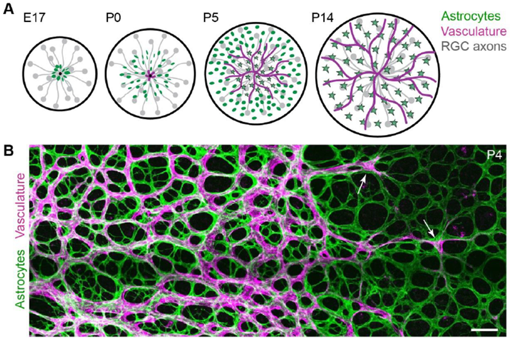
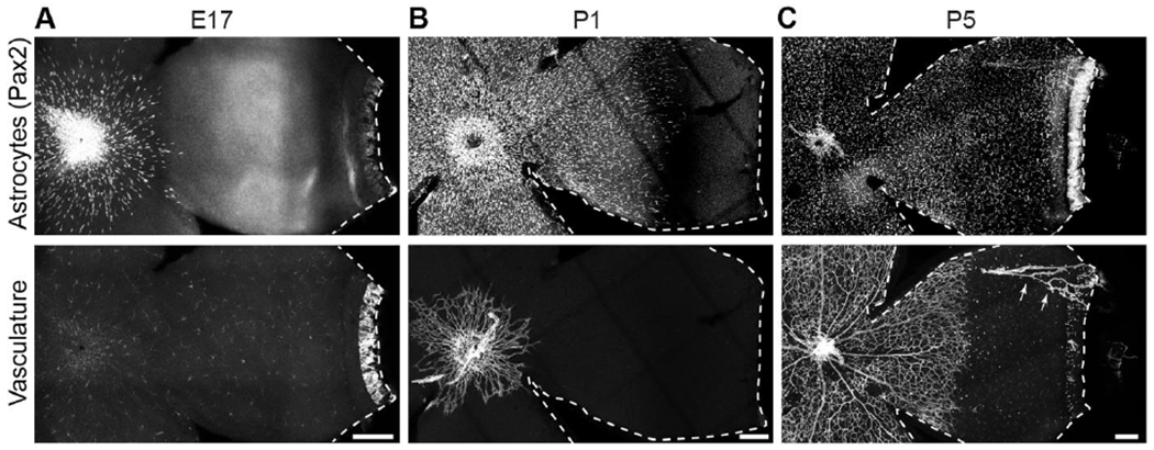
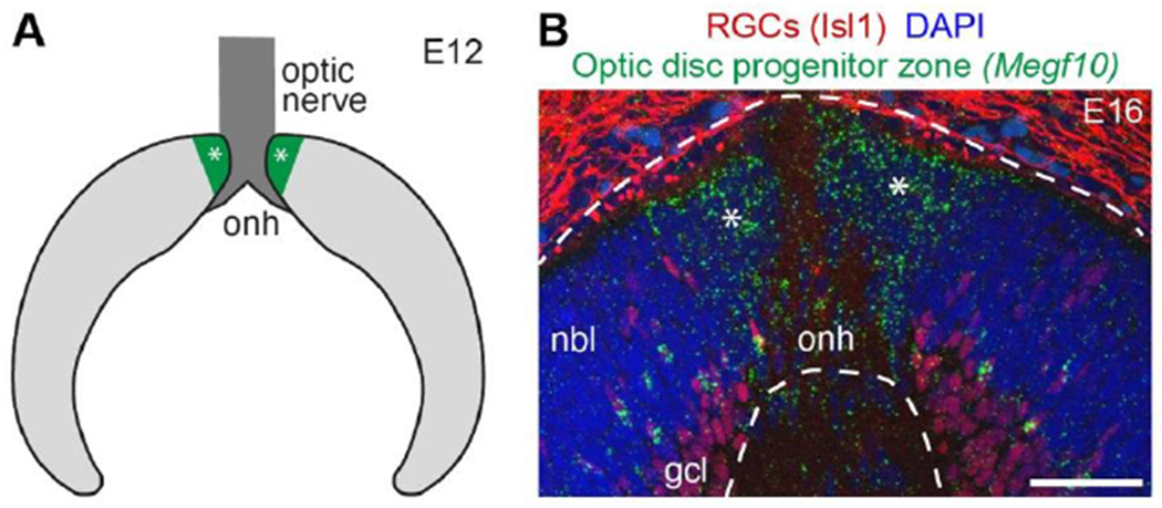
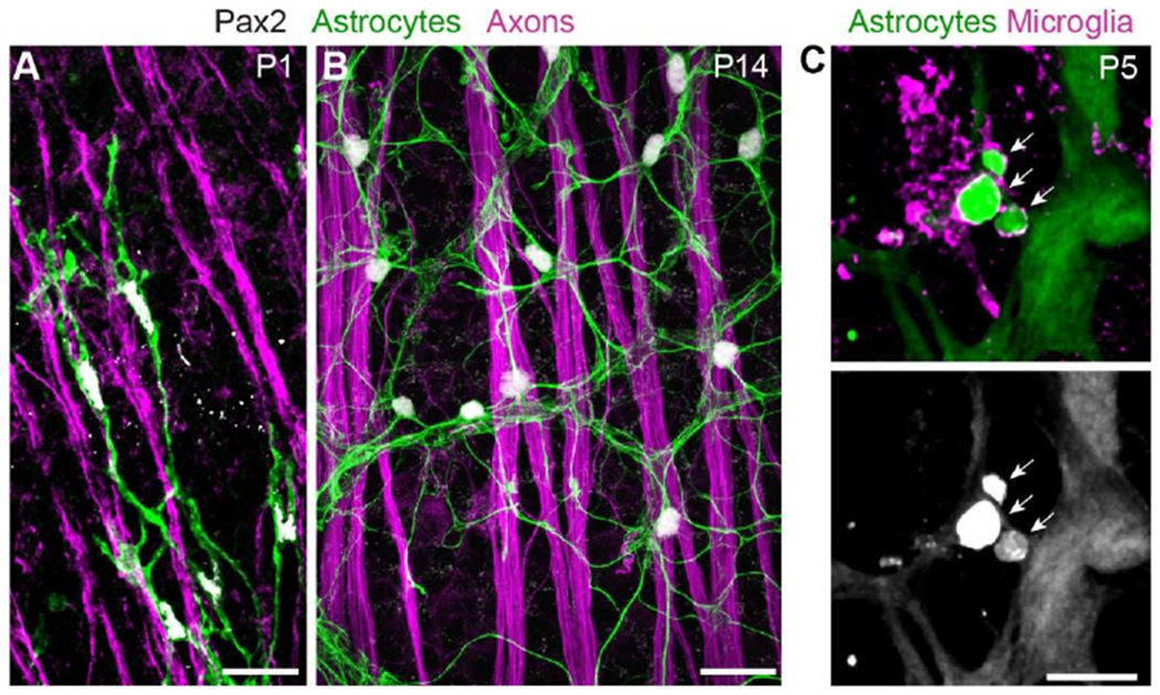
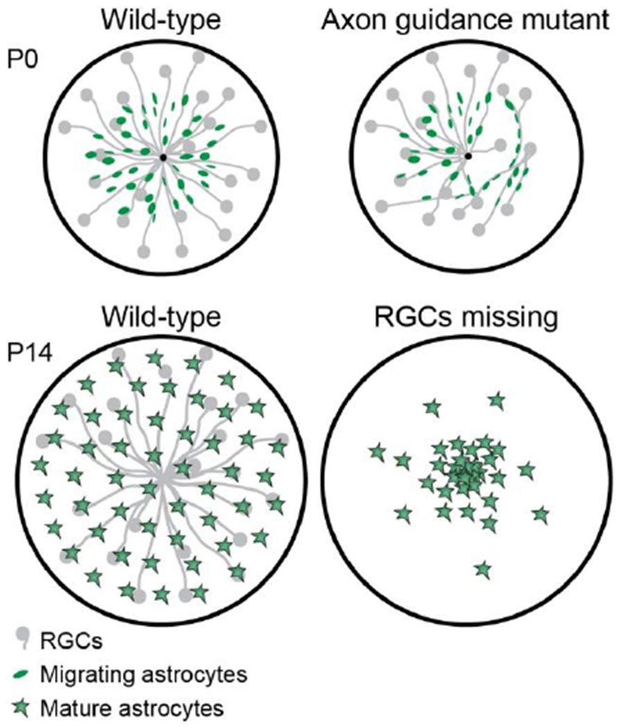
Throughout the central nervous system, astrocytes adopt precisely ordered spatial arrangements of their somata and arbors, which facilitate their many important functions. Astrocyte pattern formation is particularly important in the retina, where astrocytes serve as a template that dictates the pattern of developing retinal vasculature. Thus, if astrocyte patterning is disturbed, there are severe consequences for retinal angiogenesis and ultimately for vision - as seen in diseases such as retinopathy of prematurity. Here we discuss key steps in development of the retinal astrocyte population. We describe how fundamental developmental forces - their birth, migration, proliferation, and death - sculpt astrocytes into a template that guides angiogenesis. We further address the radical changes in the cellular and molecular composition of the astrocyte network that occur upon completion of angiogenesis, paving the way for their adult functions in support of retinal ganglion cell axons. Understanding development of retinal astrocytes may elucidate pattern formation mechanisms that are deployed broadly by other axon-associated astrocyte populations.

Keywords: Angiogenesis; Astrocyte; Hypoxia-inducible factor; Microglia; PDGF; Retinal development; Retinal ganglion cells; Retinopathy of prematurity; VEGF; Vascular development.


Conflict of interest statement

Declaration of Interests

The authors declare no competing interests.

Figures

Figure 1:
Figure 1:. Development of the retinal nerve fiber layer.
A) Schematic detailing sequential development of cell types comprising the RNFL. Before birth (E17 in mice), astrocytes begin to migrate into the RNFL using RGC axons as guides. Vessels enter postnatally (P0). Astrocytes mature as vessels pass over (stars, mature astrocytes). B) En face confocal photomicrograph of flat-mounted retina depicting RNFL angiogenesis. Incoming vasculature (magenta; labeled by Griffonia simplicifolia Isolectin B4) progresses towards peripheral retina (at right), directly following the preformed astrocyte network (green, anti-PDGFRα). Arrows indicate tip cells at the angiogenic vanguard. Scale bar, 100 μm. (A) modified from Puñal et al. (2019); (B) modified from Perelli et al. (2021).
Figure 2:
Figure 2:. Migration of retinal astrocytes precedes angiogenesis.
Flat-mounted mouse retinas labeled with markers of astrocyte nuclei (Pax2) and vasculature (A,C: Isolectin B4; B: anti-CD31). A) Astrocytes begin to enter the retina before birth (E17), prior to endothelial cell entry. No vasculature is present although lectin-labeled microglia are visible. B) By P1, astrocytes cover over half of the RNFL. Vasculature is just beginning to exit the optic nerve head. C) At P5, astrocytes have reached the periphery and vasculature covers approximately half of the retina. Arrows, hyaloid vessels that were not completely removed during retinal dissection. Ahead of the angiogenic wavefront astrocyte somata are arranged in a circular pattern, mirroring astrocyte arbors (Fig. 1B). Dotted lines delineate edge of retinal tissue. Scale bars, 100 μm.
Figure 3:
Figure 3:. Retinal astrocytes are produced by specialized neuroepithelial progenitors.
A) Schematic depicting cross-section of embryonic mouse retina. The optic disc progenitor zone (green, asterisks), which gives rise to retinal astrocytes, surrounds the optic nerve head (dark gray). Light gray, retinal progenitors derived from the optic cup; these give rise to retinal neurons and Müller glia. B) Photomicrograph of E16 mouse retinal cross section. In situ hybridization for Megf10 gene, an astrocyte marker, reveals location of optic disc progenitor zone (asterisks). Dashed lines demarcate neural retina. Anti-Isl1 staining shows location of RGC cell bodies (also note non-specific staining outside neural retina). Abbreviations: onh, optic nerve head; nbl, neuroblast layer containing retinal progenitors; gcl, ganglion cell layer. Scale bar, 50 μm.
Figure 4:
Figure 4:. Cell-cell interactions of retinal astrocytes.
A,B) Interactions with axons (labeled by anti-Neurofilament medium chain). At P1 (A), astrocyte precursor cells climb along RGC axons as they migrate towards peripheral retina. Note polarization of astrocyte arbors (revealed by Pax2-Cre-driven membrane-GFP reporter) along the axon bundles. At P14 (B), mature astrocytes are no longer closely associated with RGC axons, instead showing uniform “mosaic” spacing. Astrocyte arbors labeled by anti-GFAP. See O’Sullivan et al. (2017) for further details on astrocyte-axon interactions during migration. C) Interactions with microglia. At P5, astrocyte debris (arrows) can be found within microglia. Astrocytes were labeled with GFAP-Cre-driven TdTomato reporter; microglia were labeled with a combination of antibodies to Iba1 and P2Y12. At this age, GFAP-Cre is selective for astrocytes and not yet expressed by Müller glia. Scale bars: 25μm (A,B), 10μm (C). (C) modified from Puñal et al. (2019).
Figure 5:
Figure 5:. Retinal ganglion cell axons guide astrocyte migration.
Schematic illustrating effects of RGC axon manipulations on astrocyte migration. Top: In retinas lacking both Robo1 and Robo2 genes, axon navigation is disrupted. Instead of projecting directly to the optic nerve head, as in wild-type retina (left), many mutant axons take meandering routes. Migrating astrocytes follow mistargeted axons regardless of their trajectories. Bottom: Atoh7 mutant mice lack RGCs. In these mutants, astrocytes can migrate a short distance from their origin but fail to reach peripheral retina. As a result their density in central retina is abnormally high. For further details see O’Sullivan et al. (2017).

References

    1. Allen NJ, Eroglu C, 2017. Cell Biology of Astrocyte-Synapse Interactions. Neuron 96, 697–708. 10.1016/j.neuron.2017.09.056 - DOI - PMC - PubMed
    1. Bosze B, Suarez-Navarro J, Soofi A, Lauderdale JD, Dressler GR, Brown NL, 2021. Multiple roles for Pax2 in the embryonic mouse eye. Dev. Biol 472, 18–29. 10.1016/j.ydbio.2020.12.020 - DOI - PMC - PubMed
    1. Bucher F, Stahl A, Agostini HT, Martin G, 2013. Hyperoxia causes reduced density of retinal astrocytes in the central avascular zone in the mouse model of oxygen-induced retinopathy. Mol. Cell. Neurosci 56, 225–33. 10.1016/j.mcn.2013.06.001 - DOI - PubMed
    1. Bugiani M, Vuong C, Breur M, van der Knaap MS, 2018. Vanishing white matter: a leukodystrophy due to astrocytic dysfunction. Brain Pathol 28, 408–421. 10.1111/bpa.12606 - DOI - PMC - PubMed
    1. Büssow H, 1980. The astrocytes in the retina and optic nerve head of mammals: a special glia for the ganglion cell axons. Cell Tissue Res 206, 367–78. 10.1007/BF00237966 - DOI - PubMed

Publication types

MeSH terms

LinkOut - more resources