. 2021 Aug;69(8):495-509.
doi: 10.1369/00221554211032008. Epub 2021 Jul 14.

Expression of Transcription Factor CREM in Human Tissues

Affiliations

Expression of Transcription Factor CREM in Human Tissues

Heidi Kaprio et al. J Histochem Cytochem. 2021 Aug.

Abstract

Cyclic AMP element modulator (CREM) is a transcription factor best known for its intricate involvement in spermatogenesis. The CREM gene encodes for multiple protein isoforms, which can enhance or repress transcription of target genes. Recent studies have identified fusion genes, with CREM as a partner gene in many neoplastic diseases. EWSR1-CREM fusion genes have been found in several mesenchymal tumors and in salivary gland carcinoma. These genes encode fusion proteins that include the C-terminal DNA-binding domain of CREM. We used a transcriptomic approach and immunohistochemistry to study the expression of CREM isoforms that include DNA-binding domains across human tissues. We found that CREM protein is widely expressed in almost all normal human tissues. A transcriptomic analysis of normal tissues and cancer showed that transcription of CREM can be altered in tumors, suggesting that also wild-type CREM may be involved in cancer biology. The wide expression of CREM protein in normal human tissues and cancer may limit the utility of immunohistochemistry for identification of tumors with CREM fusions.

Keywords: CREB; CREM; ICER; cancer; cyclic AMP element modulator; fusion gene; immunohistochemistry; normal tissue; transcription factor.


Conflict of interest statement

Competing Interests: The author(s) declared no potential conflicts of interest with respect to the research, authorship, and/or publication of this article.

Figures

Figure 1.
Figure 1.
Schematic presentation of CREM gene, CREM/ICER proteins, and antibody target sequence. Exon structure of the CREM gene showing CREM alternatively used promoters P1, P3, P4, and ICER promoter P2. Arrows indicate the start of transcription. ATG indicates the start of translation and STOP codons. Exon size and intron distance are not to scale. The corresponding functional domains of CREM and ICER protein are noted below. The transactivation domains consist of exons E and F encoding the regulated phosphorylation [P-box; also called kinase-inducible domains (KID)] and the two glutamine-rich (Q-rich) exons involved in basal transactivational activities, which are absent in ICERs. The DNA-binding domains are exons H and I that encode the basic regions required for DNA recognition and the leucine zipper dimerization domains, respectively. Exon I can be alternatively spliced in Ia and Ib. Antigenic sequence targets amino acids 201–300 of protein sequence, corresponding to exons H and I, that includes DNA-binding domain. Abbreviations: CREM, cyclic AMP element modulator; ICER, inducible cAMP early repressor.
Figure 2.
Figure 2.
Validation of the antibody used for immunohistochemical stainings using Western Blotting and siRNA-mediated knockdown of CREM expression. Western blotting using cell lines CHL-1, HEK-239, and PC-3 shows bands of expected size using the CREM antibody (between 20 and 37 KDa depending on isoform). After transfection using CREM-targeting siRNA, the bands are markedly weaker, indicating that the knockdown of CREM is successful and that the antibody specifically detects CREM (blue bands). The bands are of different size than those seen using CREB (expected size of 43 KDa; data not shown). After CREM siRNA treatment, only bands detected by anti-CREM are weaker, confirming that this antibody detects CREM specifically. EWSR1-CREM fusion (≈55 KDa) in the CHL-1 cell line was detected with both anti-CREM and EWSR1 antibodies (superpositions of blue and green). Endogenous EWSR1 (green bands) was not affected by CREM knockdown, and GAPDH shows equal amounts of protein loading. Molecular marker: Dual Color Precision Plus Protein standards (Bio-Rad). Abbreviations: CREM, cyclic AMP element modulator; GADPH, glyceraldehyde-3-phosphate dehydrogenase; siRNA, small interfering RNA.
Figure 3.
Figure 3.
Analysis of CREM mRNA expression in different tissues using the GTEX database. Expression of CREM mRNA in different human tissues, arranged from the highest expression to the lowest expression. Values are shown in TPM (transcripts per million). No other normalization steps have been applied. Abbreviations: CREM, cyclic AMP element modulator.
Figure 4.
Figure 4.
Immunohistochemical detection of CREM in human tissues. (A) The four-tier immunohistochemical evaluation scheme for CREM stainings: 0 negative (cerebellum), 1 weak (pancreas), 2 moderate (stomach), and 3 strong (parathyroid). Scale bar = 20 µm. (B) In the cerebellum, we detected virtually no nuclear staining. Black arrow indicates a Purkinje cell on the border of the molecular and granular cell layers. (C) In the respiratory epithelium of the nasal cavity, 60–69% of cells were positive with dominantly moderate staining intensity. The negative cells were usually of goblet cell type. (D) The palate squamous epithelium had a gradient expression profile. (E) The apical cells (AC) of the gastric corpus were negative, whereas the glandular cells expressed CREM. (F) The duodenal stromal cells had weak to moderate staining intensity, whereas enterocytes and goblet cells were predominantly negative near the crypts as seen here. Apical enterocytes had more CREM expression. (G) In the submandibular gland, the mucous glands (MG) were weaker and more narrow in their CREM expression of the ducts (D) and serous glands (SG). (H) The hepatocytes of the liver had negative nuclei and a moderate granular cytoplasmic staining. (I) In the kidney glomerulus (G), only just over half of the nuclei stained positive, and the proximal tubules (PT) and distal tubules (DT) had wider expression. (J) In the urinary bladder epithelium, most of the nuclei had CREM expression. (K) The ciliated columnar cells of the fimbriae epithelium were among the strongest staining cell types. (L) In the placenta, the tertiary villi had negative syncytiotrophoblasts lining the villi, whereas half of the cytotrophoblasts had moderate CREM expression. (M) In the testis, seminiferous tubules (T) had different nuclear staining intensities, from negative to strong. (N) The white pulp (W) in the spleen was mostly negative, whereas in the red pulp (R) we observed clear staining of the sinusoidal cell nuclei. (O) In the lymph node cortex, the germinal center (G) had a near-uniform positive CREM expression, whereas interestingly the mature cortical lymphocytes (M) remained almost negative. (P) Skeletal muscle nuclei had a wide weak to moderate staining pattern. Here, the capillaries and supporting tissue are nearly negative (C). The scale bar in picture O is 20 µm; all images are of the same magnification. Images of the 15 tissues in B–P in smaller magnification are available as supplements. Abbreviation: CREM, cyclic AMP element modulator.
Figure 5.
Figure 5.
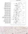
Transcriptomic analysis of CREM in cancer and immunohistochemical staining in mucoepidermoid carcinoma. (A) CREM mRNA expression in human normal tissues and cancer. The logarithmic expression values (log2) of CREM are presented on the x-axis as box-plots. The box extends from the first to the third quartile, and the median is presented as a line. The whiskers extend to data points that exist within 1.5× interquartile range (IQR) lower than the first quartile or 1.5× IQR higher than the third quartile. Beyond these values, data points were interpreted as outliers; mild outliers are marked with a circle, whereas extreme outliers are marked with asterisk. Two thirds of the normal tissue versus cancer pairs selected here have downregulation of CREM in cancer tissue; in the rest of the pairs, the corresponding cancer tissues have CREM overexpression. Values of p from Student’s t-test are indicated. (B) Mucoepidermoid carcinoma with an EWSR1-CREM fusion stained with anti-CREM. Moderate positive staining can be seen homogeneously in a majority of tumor cell nuclei. (C) CREM fusion–negative mucoepidermoid carcinoma stained with anti-CREM. Also here, CREM is positive in most cell nuclei at moderate intensity. Scale bar, 50 µm. Abbreviations: CREM, cyclic AMP element modulator.

References

    1. Brindle PK, Montminy MR. The CREB family of transcription activators. Curr Opin Genet Dev. 1992;2(2):199–204. - PubMed
    1. Sassone-Corsi P. Coupling gene expression to cAMP signalling: role of CREB and CREM. Int J Biochem Cell Biol. 1998;30(1):27–38. - PubMed
    1. Laoide BM, Foulkes NS, Schlotter F, Sassone-Corsi P. The functional versatility of CREM is determined by its modular structure. EMBO J. 1993;12(3):1179–91. - PMC - PubMed
    1. Kosir R, Juvan P, Perse M, Budefeld T, Majdic G, Fink M, Sassone-Corsi P, Rozman D. Novel insights into the downstream pathways and targets controlled by transcription factors CREM in the testis. PLoS One. 2012;7(2):e31798. - PMC - PubMed
    1. Wu X, Jin W, Liu X, Fu H, Gong P, Xu J, Cui G, Ni Y, Ke K, Gao Z, Gao Y. Cyclic AMP response element modulator-1 (CREM-1) involves in neuronal apoptosis after traumatic brain injury. J Mol Neurosci. 2012;47(2):357–67. - PubMed

Publication types

MeSH terms

Substances