. 2021 Jul 20;118(29):e2106203118.
doi: 10.1073/pnas.2106203118.

In vitro affinity maturation of broader and more-potent variants of the HIV-1-neutralizing antibody CAP256-VRC26.25

Affiliations

In vitro affinity maturation of broader and more-potent variants of the HIV-1-neutralizing antibody CAP256-VRC26.25

Yiming Yin et al. Proc Natl Acad Sci U S A. .

Abstract

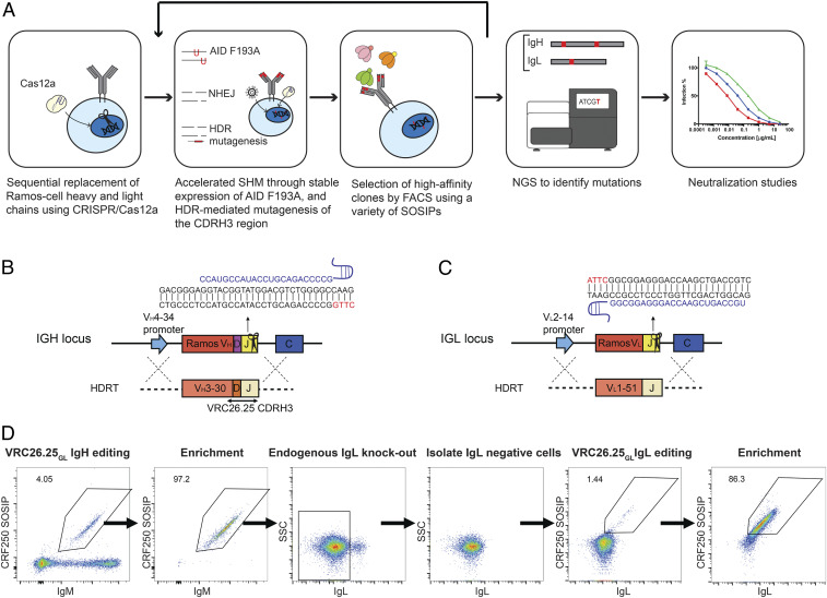
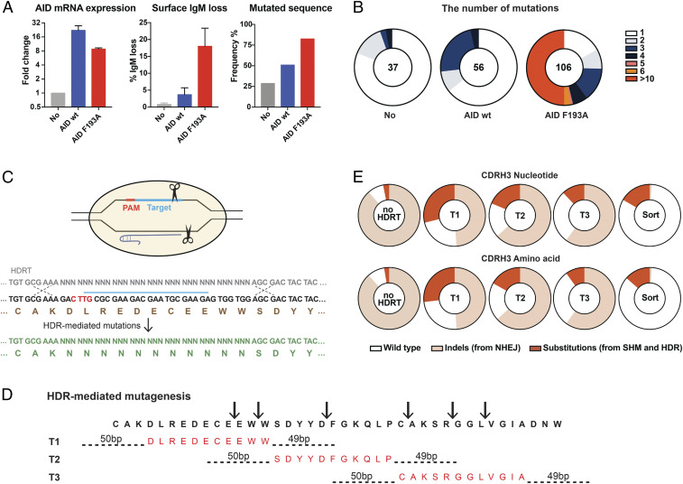
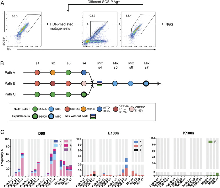
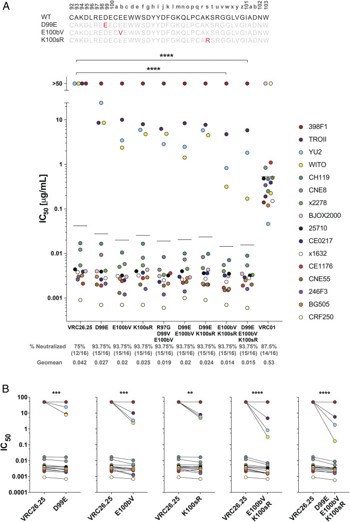
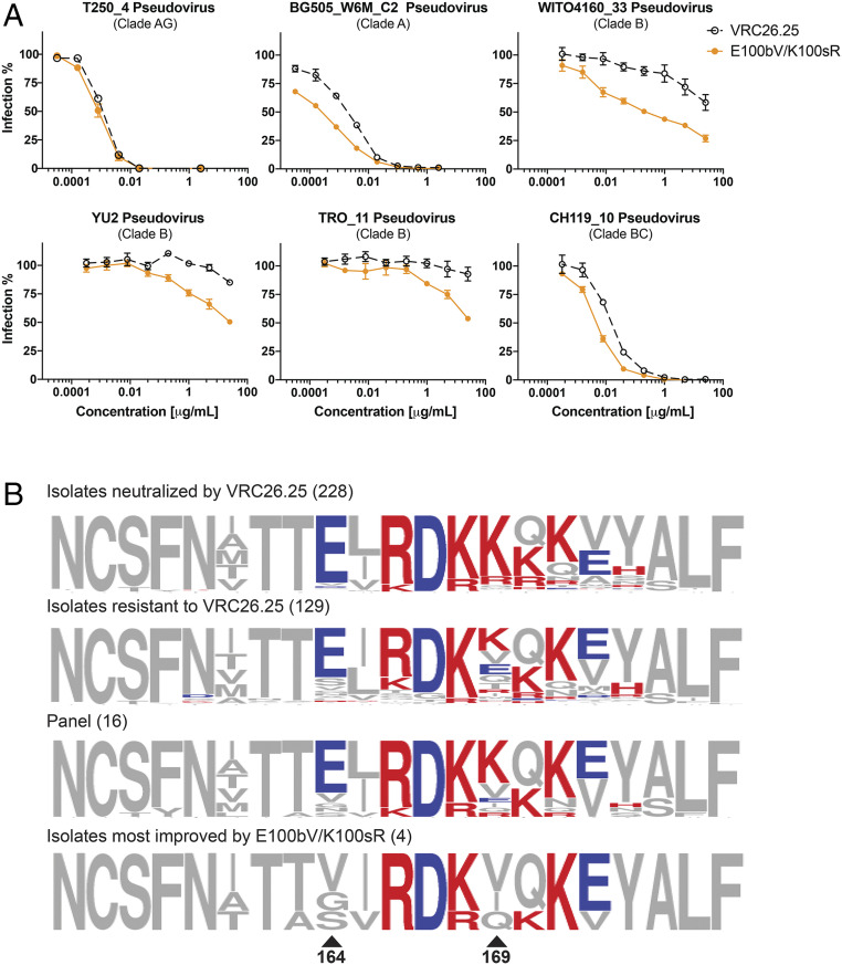
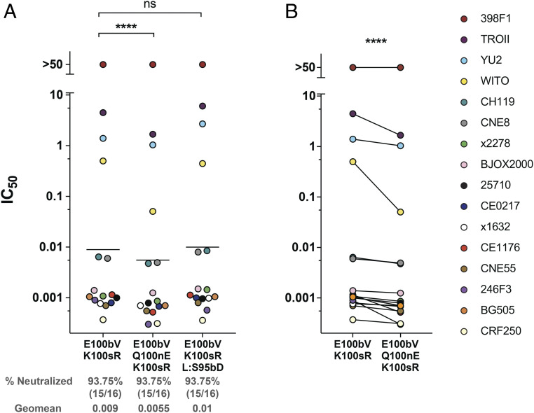
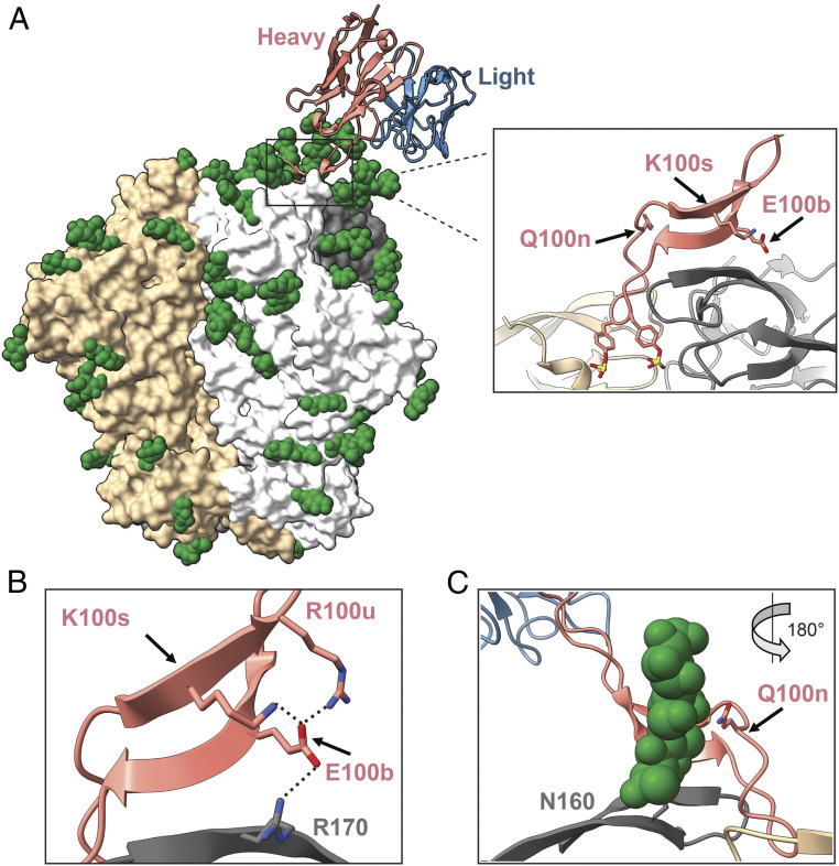
Three variable 2 (V2) loops of HIV-1 envelope glycoprotein (Env) trimer converge at the Env apex to form the epitope of an important classes of HIV-1 broadly neutralizing antibodies (bNAbs). These V2-glycan/apex antibodies are exceptionally potent but less broad (∼60 to 75%) than many other bNAbs. Their CDRH3 regions are typically long, acidic, and tyrosine sulfated. Tyrosine sulfation complicates efforts to improve these antibodies through techniques such as phage or yeast display. To improve the breadth of CAP256-VRC26.25 (VRC26.25), a very potent apex antibody, we adapted and extended a B cell display approach. Specifically, we used CRISPR/Cas12a to introduce VRC26.25 heavy- and light-chain genes into their respective loci in a B cell line, ensuring that each cell expresses a single VRC26.25 variant. We then diversified these loci through activation-induced cytidine deaminase-mediated hypermutation and homology-directed repair using randomized CDRH3 sequences as templates. Iterative sorting with soluble Env trimers and further randomization selected VRC26.25 variants with successively improving affinities. Three mutations in the CDRH3 region largely accounted for this improved affinity, and VRC26.25 modified with these mutations exhibited greater breadth and potency than the original antibody. Our data describe a broader and more-potent form of VRC26.25 as well as an approach useful for improving the breadth and potency of antibodies with functionally important posttranslational modifications.

Keywords: B cell display; CAP256-VRC26.25; V2-glycan bNAbs; affinity maturation; tyrosine sulfation.


Conflict of interest statement

The authors declare no competing interest.

Figures

Fig. 1.
Fig. 1.
Introduction of VRC26.25GL heavy- and light-chain genes into Ramos cells. (A) The workflow of these studies is represented. The heavy- and light-chain loci of Ramos cells were edited to encode VRC26.25GL. These loci were then diversified through stable expression of an AID variant (F193A) and through libraries of HDRTs targeting the CDRH3 region. BCRs were iteratively selected with various SOSIP trimers, and the B cell loci of selected cells were deep sequenced after each selection. Enriched mutations were introduced into mature, wild-type VRC26.25, and the resulting bNAb variants were assayed for their ability to neutralize a panel of global HIV-1 isolates. (B) A strategy for replacing the variable region of the Ramos-cell heavy chain. Cas12a recognizing a CTTG PAM region (red) cleaved a region encoded by the JH6 gene. Sequences of the single-stranded Cas12a gRNA and the double-stranded target are shown. The native gene was replaced using an HDRT encoding a 5′ homology arm that complements the V4-34 promoter, followed by the V3-30 leader sequence with intron, the VRC26.25GL heavy-chain variable region, and a 3′ homology arm that complements the intron after JH6, as indicated. (C) Similarly, the Ramos lambda light-chain locus was cleaved in a region encoded by the JL2 gene, and the native light-chain gene was replaced using an HDRT encoding a 5′ homology arm complementing the V2-14 promoter, the V1-51 leader with intron, the VRC26.25GL light-chain, and a 3′ homology arm that complements the intron after JL2. (D) The process of replacing the native Ramos antibody genes with the VRC26.25GL heavy and light chains. The heavy chain was introduced first using the approach shown in panel B. Heavy chain–edited cells were then enriched by FACS using the SOSIP protein based on the CRF250 isolate (CRF250 SOSIP, vertical axis), and an anti-IgM antibody (IgM, horizontal axis). Expression of the native Ramos light-chain was eliminated by NHEJ, and the VRC26.25GL light chain was introduced using the approach shown in panel C, rescuing BCR expression in successfully edited cells. Finally, VRC26.25GL BCR-expressing cells were again enriched by FACS using CRF250 SOSIP and IgL antibodies.
Fig. 2.
Fig. 2.
Accelerated BCR diversification in Ramos cells. (A) Ramos cells were transduced with retroviruses expressing AID or an AID variant (F193A). AID expression was measured by qRT-PCR assay 2 wk posttransduction. Expression is shown normalized to the endogenous AID expressed in untransduced cells. The error bars represent SD of triplicates (Left). Cell-surface IgM expression was measured by flow cytometry 2 wk post transduction. The bars represent loss of IgM expression, an indication of AID activity. The error bars represent SD of two independent experiments (Center). The percentage of mutated heavy chain, determined by NGS 1 mo posttransduction, was compared in unmodified Ramos cells, where a baseline level of mutation is observed, and in cells transduced with AID or AID F193A (Right). (B) An analysis of NGS results in which the number of mutations per sequence analyzed is presented. The number of analyzed sequences with >100 UMI are shown in the Center of each chart. (CE) In addition to AID-mediated mutations, the heavy chains of AID F193A–transduced VRC26.25GL cells were diversified through HDR-mediated randomization, as shown. (C) An example of HDRT-mediated random mutagenesis is presented. After Cas12a-mediated cleavage, soft-randomized HDRT introduced mutations into the VRC26.25 CDRH3. “N” represents 91% original wild-type bases with 3% of each of the other nucleotides. (D) The strategy used to randomize the full VRC26.25 CDRH3 is shown. The CDRH3 region was cleaved at six locations using Cas12a and six distinct gRNAs, as indicated by arrows. One of three HDRTs (T1, T2, and T3), each soft randomized in an 11– or 12–amino acid region (red), was used to edit the cleaved CDRH3 target. HDRT homology arms are indicated by dotted lines. (E) Pie charts represent the proportion of indels and substitutions in the CDRH3 of HDRT-mutated cells, as determined by NGS after one round of editing in AID F193A–transduced VRC26.25GL cells. These cells were electroporated with RNP including all six gRNAs, in the absence (no HDRT) or presence of the indicated HDRT. Cells mutated using HDRT T1, T2, and T3 were combined, sorted with CRF250 SOSIP and an anti-IgL antibody, and again analyzed (sort). Nucleotide (Top) and amino acid (Bottom) changes are shown.
Fig. 3.
Fig. 3.
Affinity maturation of VRC26.25 in vitro. (A) VRC26.25GL-expressing cells sorted as shown in Fig. 1D (Right panel) were diversified through the introduction of AID F193A and CRISPR-mediated editing in the CDRH3 regions. The top 0.5 to 3% SOSIP-positive IgL-positive cells were selected with a diagonal gating (Center) to reduce biases from high BCR expression. Most sorted cells bound SOSIP trimers efficiently (Right). (B) The selection cycle represented in panel A was repeated seven times for each of three pathways (Path A, B, and C), selecting with distinct sets of SOSIP antigens, as indicated with colored circles. Cells were mutated with soft-randomized HDRT and sorted four times using progressively lower concentrations of fluorescently labeled SOSIP trimers (s1 to s4). Cells were mixed (Mix-s4) and sorted an additional three cycles (Mix s5 to s7) with a SOSIP variant bearing a WITO apex region resistant to VRC26.25 (light blue circles). NGS analysis was performed after each selection step. Circles with thin boundaries represent SOSIP trimers produced from GnTI cells that lack complex glycans, whereas circles with thick boundaries indicate SOSIP molecules produced from Expi293 cells and decorated with complex glycans. (C) The bars represent the frequency of mutations at the indicated heavy-chain residues as the emerged from sort 1 (s1) to sort 7 (s7). Gray indicates the original wild-type residue.
Fig. 4.
Fig. 4.
VRC26.25 variants with improved potency and breadth. (A) The amino acid sequence of the wild-type VRC26.25 CDRH3 is shown with Kabat numbering indicated. Key mutations D99E, E100bV, and K100sR (red) are also shown aligned with the wild-type sequence. HIV-1 pseudoviruses of indicated isolates were incubated with the indicated VRC26.25 variant or the CD4-binding site bNAb VRC01 in TZM-bl cells. Measured IC50 values were plotted. Pseudoviruses resistant to the indicated antibody were assigned an IC50 of 50 µg/mL. Each dot represents the mean from two independent experiments of triplicates. A previously defined 12-isolate global panel was evaluated together with four additional isolates, namely CRF250, BG505, YU2, and WITO. Geometric mean values are indicated by horizontal lines, and their numerical values are provided beneath the figure, along with the percent of 16 isolates neutralized by the antibody-indicated variant. (B) IC50 values of the indicated VRC26.25 variant was compared to the IC50 of wild-type VRC26.25 for each of the 16 isolates in the panel. Significance in both panels was determined by a Wilcoxon matched signed rank test: ns, not significant; *P < 0.05, **P < 0.01, ***P < 0.001, ****P < 0.0001.
Fig. 5.
Fig. 5.
Env determinants of enhanced VRC26.25 potency and breadth. (A) TZM-bl neutralization curves of the most potent VRC26.25 variant against pseudotyped viral isolates whose SOSIPs were used during selection (Top), and those isolates whose neutralization was most improved by these variants (Bottom; note that WITO is in both groups). Errors bars represent SEM of triplicates. (B) The amino acid alignment of susceptible and resistant viruses against VRC26.25 (first and second row), the 16-panel isolates (third row), and those isolates whose neutralization was most improve by the E100bV and K100sR mutations (fourth row) are shown. Two positions critical to viral escape from wild-type VRC26.25 are indicated with triangles. Basic and acidic amino acids were labeled in red and blue, respectively.
Fig. 6.
Fig. 6.
Further improvement of VRC26.25 variants. (A) A figure similar to that in Fig. 4A, except that IC50 values of the indicated modifications of E100bV/K100sR are compared to E100bV/K100sR itself. L preceding a mutation indicates a light-chain residue. Each dot represents the mean from two independent experiments of triplicates. Geometric means were indicated by horizontal lines. (B) A figure similar to that in Fig 4B, except that the E100bV/K100sR variant was compared to E100bV/Q100nE/K100sR variant. Significance was determined by a Wilcoxon matched signed rank test: ns, not significant; *P < 0.05, **P < 0.01, ***P < 0.001, ****P < 0.0001.
Fig. 7.
Fig. 7.
The structure of VRC26.25 in complex with CAP256.wk34.c80 SOSIP (Protein Data Bank: 6VTT). (A) The structure of a VRC26.25 Fab domain complexed with CAP256.wk34.c80 SOSIP trimer. Three SOSIP protomers are represent in gray, white, and tan surfaces, respectively. Green spheres indicate glycans resolved in the structure. The VRC26.25 heavy chain is shown in salmon, and the light chain is shown in blue. Note that the heavy-chain CDRH3 contacts all three Env protomers. Inset presents this CDRH3 with E100b, Q100n, and K100s shown and labeled. Note also the two CDRH3 sulfotyrosines (100h and 100i) that project into the apex cavity. (B) Residue E100b interacts with a network basic of residues including SOSIP R170 (gray) and VRC26.25 CDRH3 residues K100s and R100u. Potential electrostatic contacts are indicated with a dashed line. (C) VRC26.25 residue Q100n contacts a critical glycan at SOSIP N160 (green).

References

    1. Gilbert P. B., et al. ., Basis and statistical design of the passive HIV-1 antibody mediated prevention (AMP) test-of-concept efficacy trials. Stat. Commun. Infect. Dis. 9, 20160001 (2017). - PMC - PubMed
    1. Corey L.et al. .; HVTN 704/HPTN 085 and HVTN 703/HPTN 081 Study Teams , Two randomized trials of neutralizing antibodies to prevent HIV-1 acquisition. N. Engl. J. Med. 384, 1003–1014 (2021). - PMC - PubMed
    1. Gautam R., et al. ., A single injection of anti-HIV-1 antibodies protects against repeated SHIV challenges. Nature 533, 105–109 (2016). - PMC - PubMed
    1. Doria-Rose N. A.et al. .; NISC Comparative Sequencing Program , Developmental pathway for potent V1V2-directed HIV-neutralizing antibodies. Nature 509, 55–62 (2014). - PMC - PubMed
    1. Walker L. M.et al. .; Protocol G Principal Investigators , Broad and potent neutralizing antibodies from an African donor reveal a new HIV-1 vaccine target. Science 326, 285–289 (2009). - PMC - PubMed

Publication types

MeSH terms

Substances