. 2021 Jul 14;13(602):eabb3631.
doi: 10.1126/scitranslmed.abb3631.

Low-dose targeted radionuclide therapy renders immunologically cold tumors responsive to immune checkpoint blockade

Affiliations

Low-dose targeted radionuclide therapy renders immunologically cold tumors responsive to immune checkpoint blockade

Ravi B Patel et al. Sci Transl Med. .

Abstract

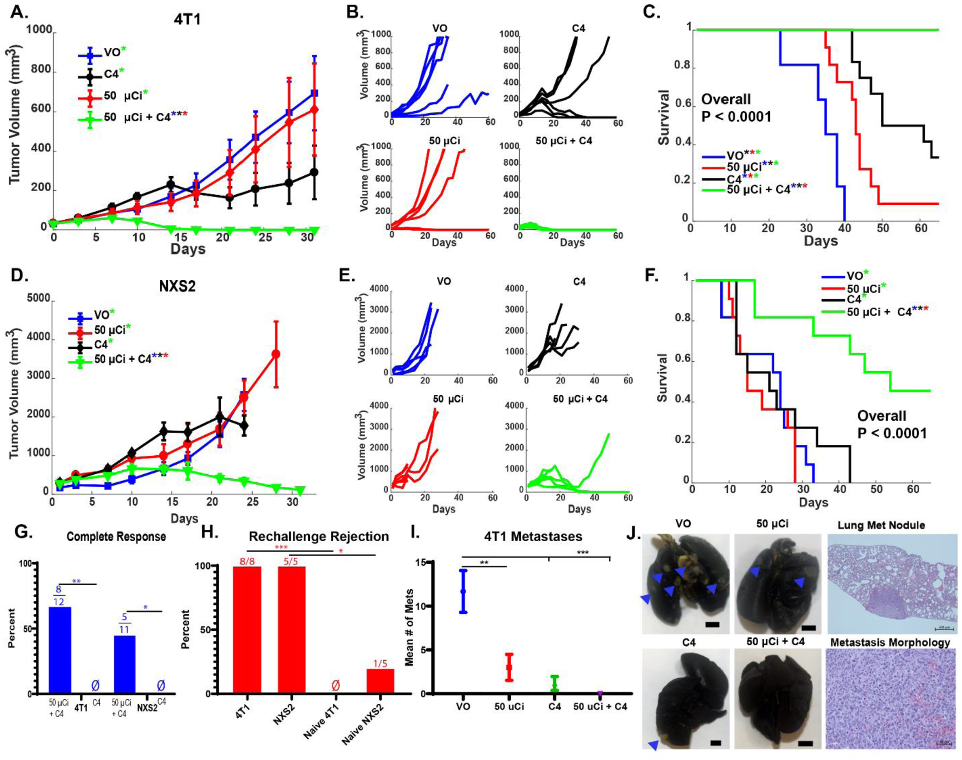
Molecular and cellular effects of radiotherapy on tumor microenvironment (TME) can help prime and propagate antitumor immunity. We hypothesized that delivering radiation to all tumor sites could augment response to immunotherapies. We tested an approach to enhance response to immune checkpoint inhibitors (ICIs) by using targeted radionuclide therapy (TRT) to deliver radiation semiselectively to tumors. NM600, an alkylphosphocholine analog that preferentially accumulates in most tumor types, chelates a radioisotope and semiselectively delivers it to the TME for therapeutic or diagnostic applications. Using serial 86Y-NM600 positron emission tomography (PET) imaging, we estimated the dosimetry of 90Y-NM600 in immunologically cold syngeneic murine models that do not respond to ICIs alone. We observed strong therapeutic efficacy and reported optimal dose (2.5 to 5 gray) and sequence for 90Y-NM600 in combination with ICIs. After combined treatment, 45 to 66% of mice exhibited complete response and tumor-specific T cell memory, compared to 0% with 90Y-NM600 or ICI alone. This required expression of STING in tumor cells. Combined TRT and ICI activated production of proinflammatory cytokines in the TME, promoted tumor infiltration by and clonal expansion of CD8+ T cells, and reduced metastases. In mice bearing multiple tumors, combining TRT with moderate-dose (12 gray) external beam radiotherapy (EBRT) targeting a single tumor augmented response to ICIs compared to combination of ICIs with either TRT or EBRT alone. The safety of TRT was confirmed in a companion canine study. Low-dose TRT represents a translatable approach to promote response to ICIs for many tumor types, regardless of location.


Conflict of interest statement

Competing interests: ZM, JW, RH, and JG have financial interest in Archeus Technologies. ZM is a member of the Scientific Advisory Boards for Archeus Technologies and for Seneca Therapeutics. PS is an unpaid Medical Advisor for Invenra Inc. JW is a co-founder, CSO, and director of Archeus Technologies which holds the license rights to NM600 related technologies. BB and JG are co-founders of Voximetry, Inc and BB is the CSO. The following patents have been applied for or filed by the University of Wisconsin Alumni Research Foundation: Pat No. US 10,736,949 Radiohalogenated Agents for in Situ Immune Modulated Cancer Vaccination with ZM, PS, JW, BB as inventors, Pat No. US 10,751,430 Targeted Radiotherapy Chelates for in Situ Immune Modulated Cancer Vaccination with ZM, PS, JW, BB, PC as inventors, App No. 15/809,427 Using targeted radiotherapy to drive anti-tumor immune response to immunotherapies – ZM, PS, JW, PC, JG, RH as inventors, and US 2011/0060602 A1 Treatment Planning System For Radiopharmaceuticals with BB and JG as inventors.

Figures

Figure 1.
Figure 1.. Imaging, uptake, and dosimetry of 86/90Y-NM600.
(A) Mice with a single (~150–250 mm3) B78, NXS2, or 4T1 flank tumor (arrow) were serially imaged using PET/CT following IV injection of 86Y-NM600. (B) Image analysis was performed to determine uptake of injected activity per gram of tissue (%IA/g) over time (at 3, 24, and 48 hr) and shows progressive accumulation of NM600 in tumor and progressive washout from normal tissues. (C) Tissue dosimetry was performed using Monte Carlo methods. (N = 3. Significance determined with one-way ANOVA with Tukey correction for multiple comparisons testing for tissue dosimetry. *P < 0.05, **P < 0.01, ***P<0.001)
Figure 2.
Figure 2.. Low-dose TRT enhances response to ICI.
(A) Mice with a single (~70–150 mm3) B78 flank tumors were treated with combinations of TRT (90Y-NM600), anti-CTLA-4 (C4), anti-PD-L1 (PDL1), or vehicle-only (VO) control injections. (B-D) Tumor response and animal survival after treatment with TRT and ICI are shown as a function of TRT dose. The mean tumor growth for each group is shown in B, the growth curves for each individual mouse in each group are shown in C and the survival for all groups in D. (E-G) Tumor response and survival based on ICI timing in relation to TRT are shown for tumor growth by group (E), by individual mouse (F), and survival by group (G). (H-J). While these data suggest comparable effect of day 4 single dose and our three-dose regimen, we have used the three-dose regimen as a standard so that our data can be evaluated in the context of prior studies that have used this regimen together with EBRT (8, 48). Tumor growth and survival for single vs dual ICI are shown for tumor growth by group (H), by individual mouse (I), and survival by group (J). In this tumor model, response and survival after treatment with anti-PD-L1 or combined anti-CTLA-4 and anti-PD-L1 is not significantly (p = ns) different from that with VO. In mice with CR to treatment in the TRT dose response studies (K), C4 timing response studies (L), and dual checkpoint studies (M), we tested for immunologic memory to B78 re-challenge and for those mice that rejected the B78 re-challenge, we then tested for rejection of subsequent B16 and Panc02 challenge as shown (K-M, Ø indicates no mice rejected challenge with symbol color representing challenge type). (N = 6 per replicate, 2 replicates). Significance determined by linear mixed effects regression analysis and two-way ANOVA with Tukey multiple comparisons testing for tumor growth [significant differences, p < 0.05, demarcated by * with the color of the asterisk representing which group from which the sample is significantly different], Kaplan–Meier with Log-rank testing for survival analysis, Chi-square contingency testing for immune memory shown in Table S1–3. * P < 0.05)
Figure 3.
Figure 3.. TRT enhances efficacy of ICI in 4T1 breast cancer and NXS2 neuroblastoma models.
Mice with a single (~70–150 mm3) 4T1 or NXS2 flank tumor were treated with VO, 50 μCi TRT (90Y-NM600), anti-CTLA-4 (C4), or 50 μCi TRT + C4. Tumor response, by group, by individual animal, and animal survival are shown for 4T1 (in A, B and C) and for NXS2 (in D, E and F). Mice with complete response to treatment with TRT + C4 (G, Ø indicates no complete responders) to initial treatment were rechallenged with the same tumor they initially rejected, and rejection rates were compared to those for naïve controls (H, Ø indicates no rejection). The 4T1 model develops spontaneous lung metastases and the number of discrete metastases at Day 25 was compared by treatment group (I). (N = 6 per replicate, 12 total). Representative lungs are shown by gross examination for the 4 treatment groups shown in I, to demonstrate what the 4T1 metastases look like in India-ink injected lungs (with blue arrows indicating 4T1 nodules that are visible by gross inspection; along with a low power photomicrograph (power of 50X) to show a lung metastases at low power, and a higher power photomicrograph (power of 400X), to show the tumor morphology within one such 4T1 nodule (J). [Significance determined by linear mixed effects regression analysis with Tukey multiple comparisons testing for tumor growth (significant differences, p < 0.05, demarcated by * with the color of the asterisk representing which group from which the sample is significantly different), Kaplan–Meier with Log-rank testing and Cox Regression for survival analysis, Chi-square contingency testing for complete response rate and immune memory, and a two-way ANOVA for number of metastases. *P < 0.05, **P < 0.01, ***P<0.001]
Figure 4.
Figure 4.. Tumor targeted radiation is necessary for enhanced efficacy of RT + ICI.
(A) C57Bl/6 mice with a single (~90 mm3) B78 tumor were treated with ~2.5 Gy RT delivered via tumor directed EBRT, whole mouse (WM) EBRT, or TRT (90Y-NM600) either alone or with anti-CTLA-4 (C4). (B-D) Tumor response was tracked for 30 days. (E) To test the effects of T cell depletion on tumor response, anti-CD4 and anti-CD8 depleting antibodies or control rat IgG were administered during TRT + C4 treatment and compared to VO (no TRT or C4 or depleting antibody) control. (F) Tumor response to 90Y-NM600 + C4 vs cold (non-radioactive) NM600 + C4 compared to VO (no NM600 or C4) control is shown. (N = 6 per replicate, 12 total. Significance determined by linear mixed effects regression analysis with Tukey multiple comparisons [significant differences, p < 0.05, demarcated by * with the color of the asterisk representing which group from which the sample is significantly different].
Figure 5.
Figure 5.. Dose and time dependent radiation effects on the TME.
Flow cytometry analyses of tumor immune cell infiltrates [total immune cells (CD45+), myeloid cells (CD11b+), T effector cells (CD8+), Tregs (CD4+CD25+FOXP3+), and NK cells (NK1.1+)] as a percent of total live cells normalized to mean of control (VO) is shown at 1, 7, 14 days after RT administration in B78 melanoma (A-F). Gene expression of chemokines, type I IFN pathway genes, acute phase inflammatory cytokines, cell adhesion and immune activation genes, and apoptosis/DNA damage repair genes in harvested tumors is displayed as the log of the fold change in expression at 1, 7, and 14 days after RT administration in comparison for values of tumors in tumor-bearing VO treated mice, obtained at the same times (days 1, 7, and 14) (G). RT effects on D7 expression of select STING pathway genes is shown (H). 50 μCi of TRT (90Y-NM600) + anti-CTLA-4 (C4) improves survival compared to single treatment controls in a B16 WT tumors but not in B16 Tmem173−/− (STING KO) tumors (I-J). (N = 5 for flow cytometry and gene expression studies, Significance determined by two-way ANOVA; N=4–6 per replicate, 8–12 total for survival studies, Kaplan–Meier with Log-rank testing and Cox Regression for survival analysis: *P < 0.05, **P < 0.01, ***P<0.001, ****P<0.0001).
Figure 6.
Figure 6.. TRT + ICI enhances immune infiltrates in the TME.
C57Bl/6 mice with B78 tumors were treated with VO, 50 μCi TRT (90Y-NM600), anti-CTLA-4 (C4), or 50 μCi TRT + C4. (A)Tumors were harvested on Day 25 post radiation (after radioisotope reached background) and tumor immune cell infiltrates [total immune cells (CD45+), lymphocytes (CD3), T effector cells (CD8+), Tregs (CD4+CD25+FOXP3+), NK cells (NK1.1+), γδ Tcells (CD3+γδTCR+), and resident memory effector cells (CD8+CD103+)] were quantitated as a percent of total live cells via flow cytometric analysis. In addition, PD1 expression on CD8 cells was quantified by MFI. (B)A tSNE transformation was calculated for a pooled sample of all CD45+ immune cells across all treatment groups and differential staining of Pd1 staining by treatment group is shown. (C) A separate cohort of mice bearing B78 tumors were treated with the same treatment groups with double the mice in the 50 μCi TRT + C4 group. Mice in the TRT + ICI group (μCi + C4) with tumors growing at Day 25 were labeled as non-responders (NR). Cytokine and chemokine concentrations in tumor lysates were measured by multiplex immunoassay. Hierarchical clustering analysis was performed, and the assay results were displayed as a Z-score for each cytokine. (D) TCRβ sequencing of TILs demonstrated increased total and unique CDR3 chains as well as decreased D50 with TRT + ICI, but no increase in Shannon diversity index. (N = 5 for flow cytometry, multiplex, and TCR sequencing studies, Significance determined by two-way ANOVA,*P < 0.05, **P < 0.01, ***P<0.001).
Figure 7.
Figure 7.. Addition of TRT to EBRT + anti-CTLA-4 enhances primary and distant tumor response.
C57Bl/6 mice with equal sized (~120 mm3) bilateral B78 tumors were treated with 50 μCi TRT (90Y-NM600) + 12 Gy to a primary tumor (P), 12 Gy P + anti-CTLA-4 (C4), 50 μCi TRT + C4, or 12 Gy P + 50 μCi TRT +C4 (A). Tumor growth at both the primary and secondary (S) tumor site was tracked for 30 days, survival for 60 days, and all mice with CR were rechallenged at D90 with B78 and on D120 with B16/Panco2 (B-E). (N = 6 mice per group in each of 2 replicate experiments, 12 mice total per treatment]. (B) shows mean tumor growth for the primary and secondary tumors in all 4 treatment groups. (C) shows overall survival for all 4 groups. (D) primary and secondary tumor growth for all mice in one of 2 replicate experiments. (E) All mice that had CR in Figure 7D were rechallenged with B78, and those that rejected it were then rechallenged with B16 and Panc02 (Ø indicates no mice rejected challenge with symbol color representing challenge type), Significance determined by linear mixed effects regression analysis with Tukey multiple comparisons testing for tumor growth [significant differences, p < 0.05, demarcated by * with the color of the asterisk representing which group from which the sample is significantly different], Kaplan–Meier with Log-rank testing and Cox Regression for survival analysis, and Chi-square contingency testing for immune memory shown in supplemental Table S4. *P < 0.05, **P < 0.01, ***P<0.001)
Figure 8.
Figure 8.. 86Y-NM600 imaging, dosimetry, and post 86Y-NM600 treatment.
Companion canines with osteosarcoma (A) and oral melanoma (B) were imaged with 86Y-NM600. Axial images are shown at 2, 24 and 48 hours, with arrows indicating the primary (index) lesion and metastatic lesions, as well as a coronal image at 48 hours. (C,D) Dose delivered to sites of tumor vs normal tissue per GBq of injected activity was determined.

References

    1. Larkin J, Chiarion-Sileni V, Gonzalez R, Grob JJ, Cowey CL, Lao CD, Schadendorf D, Dummer R, Smylie M, Rutkowski P, Ferrucci PF, Hill A, Wagstaff J, Carlino MS, Haanen JB, Maio M, Marquez-Rodas I, McArthur GA, Ascierto PA, Long GV, Callahan MK, Postow MA, Grossmann K, Sznol M, Dreno B, Bastholt L, Yang A, Rollin LM, Horak C, Hodi FS, Wolchok JD, Combined Nivolumab and Ipilimumab or Monotherapy in Untreated Melanoma, N Engl J Med 373, 23–34 (2015). - PMC - PubMed
    1. Postow MA, Chesney J, Pavlick AC, Robert C, Grossmann K, McDermott D, Linette GP, Meyer N, Giguere JK, Agarwala SS, Shaheen M, Ernstoff MS, Minor D, Salama AK, Taylor M, Ott PA, Rollin LM, Horak C, Gagnier P, Wolchok JD, Hodi FS, Nivolumab and Ipilimumab versus Ipilimumab in Untreated Melanoma, N Engl J Med 372, 2006–2017 (2015). - PMC - PubMed
    1. Tawbi HA, Burgess M, Bolejack V, Van Tine BA, Schuetze SM, Hu J, D’Angelo S, Attia S, Riedel RF, Priebat DA, Movva S, Davis LE, Okuno SH, Reed DR, Crowley J, Butterfield LH, Salazar R, Rodriguez-Canales J, Lazar AJ, Wistuba II, Baker LH, Maki RG, Reinke D, Patel S, Pembrolizumab in advanced soft-tissue sarcoma and bone sarcoma (SARC028): a multicentre, two-cohort, single-arm, open-label, phase 2 trial, The Lancet Oncology 18, 1493–1501 (2017). - PMC - PubMed
    1. Ben-Ami E, Barysauskas CM, Solomon S, Tahlil K, Malley R, Hohos M, Polson K, Loucks M, Severgnini M, Patel T, Cunningham A, Rodig SJ, Hodi FS, Morgan JA, Merriam P, Wagner AJ, Shapiro GI, George S, Immunotherapy with single agent nivolumab for advanced leiomyosarcoma of the uterus: Results of a phase 2 study: Nivolumab for Uterine Leiomyosarcoma, Cancer 123, 3285–3290 (2017). - PMC - PubMed
    1. Lu Y-C, Robbins PF, Targeting neoantigens for cancer immunotherapy: Table 1., INTIMM 28, 365–370 (2016). - PMC - PubMed

Publication types

Substances